An orthopedic residency program uses Anatomy.tv’s 3D anatomy models to run a weekly 20-minute surgical anatomy session. Faculty rotate and isolate structures to show operative relationships that textbooks and static atlases cannot. At the program’s request, the institution and faculty are not named. The teaching methods, the tools, and the results are real, and drawn from the educator’s own account.
Why is 3D anatomy hard to teach with 2D tools?
Anatomy is a three-dimensional subject, and for too long it has been taught with two-dimensional tools. Textbooks and atlases fix the body in a few set views, usually anterior, posterior, or lateral. Students then have to rotate the structure in their heads to picture how it really sits. The research is consistent on this point. Learners struggle to build accurate spatial understanding of complex structures from textbooks, atlases, and slides alone (Triepels et al., 2020; Usmani et al., 2022).
Two pressures widen that gap. Anatomy teaching hours have fallen across many medical and health science programs over the past few decades (Memon, 2018; American College of Surgeons). And dissection-based labs, still valued for the feel of real tissue, carry real costs in money, time, expertise, storage, and access. Those costs often limit how much students can use them (Estai & Bunt, 2016).
For surgical training the limits are even sharper. A surgeon needs to see a structure from the angle of the operative approach, not the angle a textbook happened to choose. A donated body helps with that, though the tissue is stiff, hard to manipulate, and some views simply cannot be reached by hand.

How does 3D software compare to textbooks and the dissection lab?
Each tool does something the others cannot. The table below compares them across five axes that matter for surgical teaching.
| Axis | 3D digital model (Anatomy.tv) | Textbook or atlas | Dissection lab |
|---|---|---|---|
| Viewing angles | Rotate and isolate any structure from any angle | Fixed views, usually anterior, posterior, or lateral | Real 3D views, limited by tissue rigidity and hand access |
| Tissue feel | None | None | High, with real tissue and haptics |
| Imaging correlation | Linked MRI and CT | Static labeled images | No built-in imaging link |
| Repeatability | Unlimited, reset and rebuild on demand | Unlimited but static | Limited, specimens degrade and cannot reset |
| Cost and access | Web-based, any device, low cost per added user | Low cost and portable | High cost in money, time, expertise, storage, and access |
How do you teach surgical anatomy with 3D software?
Recently, the Director of Education at an institution using Anatomy.tv, a veteran orthopedic surgeon with more than two decades leading a residency, shared how it amplifies learning. They run a 20-minute anatomy session every week in a conference room. Each session walks residents through one clinically relevant view on Anatomy.tv’s 3D models. (Anatomy.tv is a web-based 3D anatomy platform from Primal Pictures. Its anatomical models are reconstructed from real human scan data and reviewed by expert anatomists.)
The program’s director rates the 3D platform above the dissection lab for building spatial understanding, while still valuing the lab for haptics training.
I think dissection labs are good for haptics learning. But truthfully, with what's going on dimensionally, Primal is more important than the anatomy lab.
- Director of Education
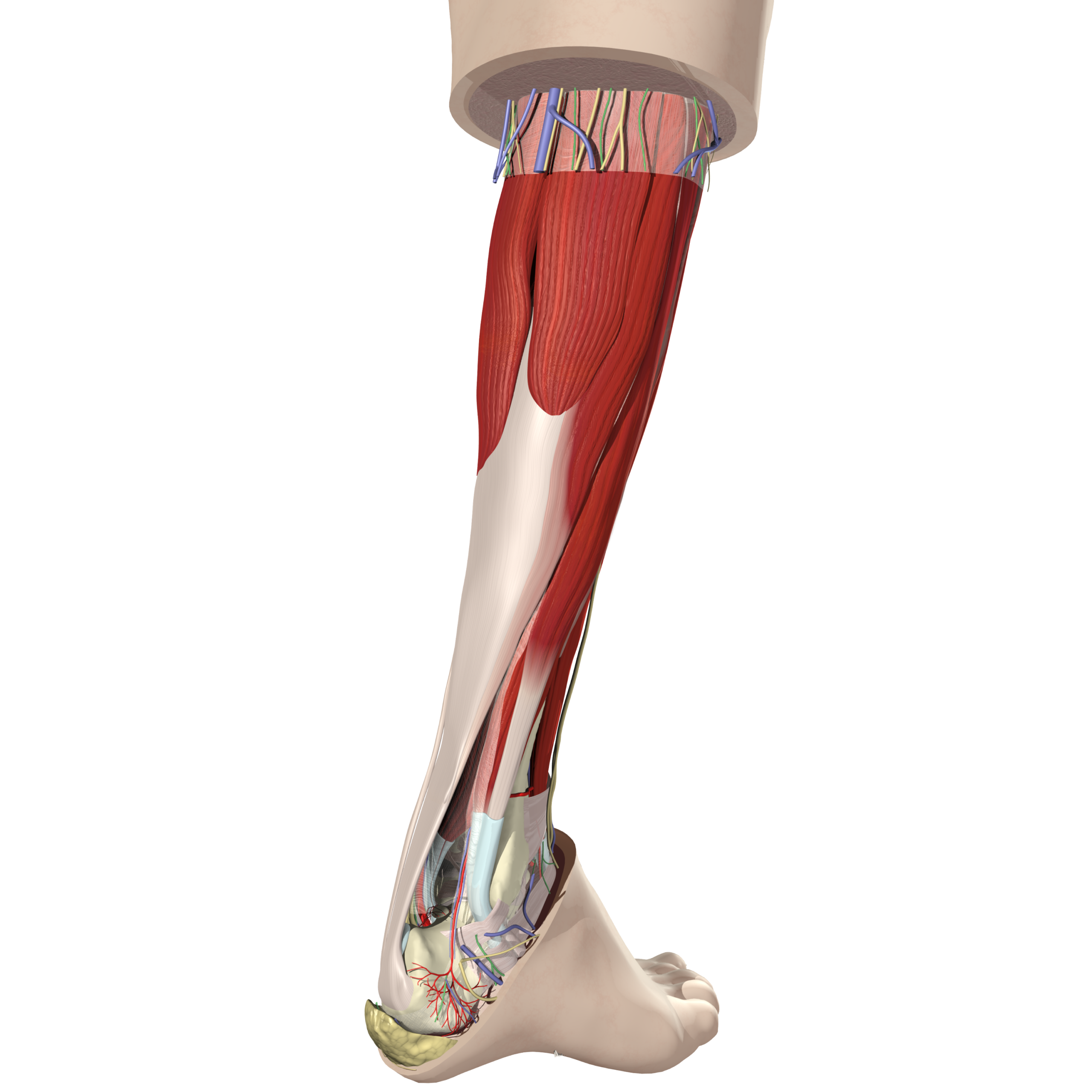
The format is simple, easy to repeat, and fits seamlessly into their current workflow and curriculum. The Director picks a structure that is hard to picture, isolates it, and rotates it so residents can see the real relationships. In one session they removed the hip anatomy down to the obturator internus and externus, and rotated to reveal the posterior aspect of the hip, where the relationship between the two muscles became clear. Another session looked at the posterolateral corner of the knee and how the capsule works with the lateral side of the knee. Rotating the model gave residents a 3D view of the muscle architecture.
How does the lateral genicular vessel integrate? What are the real relationships of the meniscus to the popliteus and the lateral collateral ligament? What you can do is give them tremendous life, because when you rotate up you now start to look from above and watch how things travel.
- Director of Education
Sessions are also tied to clinical reality. At the end of each, the Director asks residents what they will face in the operating room the following week and builds the next session around it.
Traditional anatomy lectures can lose a video-raised audience fast. A 3D digital model that moves, that residents can watch being taken apart and rebuilt, holds the room. The program moved from hand-drawn teaching slides to Anatomy.tv’s interactive 3D models.
You guys have changed our lives. From when we had to hand draw our slides to PowerPoint and then moving on, what Primal has done completely changed the game of anatomy for me. This was not the way the book was presenting it, but the way we needed it.
- Director of Education
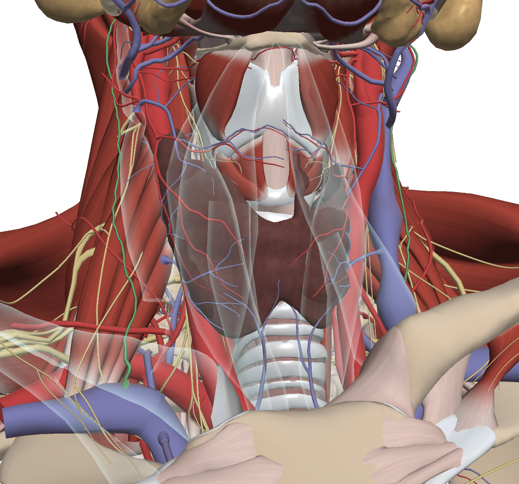
How can you use the Anatomy.tv portfolio in practice?
The platform includes several capabilities that map directly to surgical teaching.
- Rotation. For example, show a shoulder from the scapular-lateral view an orthopedic surgeon actually operates from, or look at a clavicle from three directions to plan where screws will go.
- Ghosting and hiding structures. Reduce a region to only the structures you want, then rebuild it. For example, reveal the middle genicular vessel hidden behind the popliteal artery by ghosting the artery and rotating the viewpoint, a perspective a static image cannot offer.
- Real scan data and verified detail. Primal’s model is reconstructed from real human scan and imaging data and checked by expert anatomists. That accuracy is why it can be treated as a working reference rather than an illustration.
- Cross-sectional imaging. Linked MRI and CT images connect the 3D model to the way modern radiology reads the body. This correlation helps learners connect three-dimensional anatomical relationships with their appearance in medical imaging.
- Access on any device. Web-based access means a surgeon with no computer nearby can pull up a region on a phone or tablet before surgery to walk a patient through the operation, or refer to the content afterward when drafting clinical notes.
There's anatomy that's just cool. There's anatomy that is fun to look at and difficult to understand. And when you play with this tool, it empowers you to understand the anatomy in a way you've never appreciated it before. It's like taking a trip somewhere where you use a map the first time, but you get to visualize it. And then you travel around the map a few times and all of a sudden you know exactly where you are. And if you go back, you know how to travel.
- Director of Education
Does VR improve surgical training outcomes?
Take anatomy further with Primal VR, an immersive and interactive virtual reality environment built on Anatomy.tv’s model. Students explore around and inside the body while building comprehension, engagement, and enthusiasm for learning.
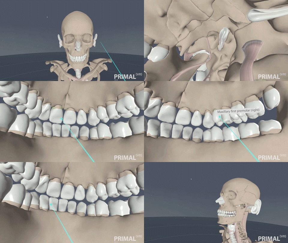
VR strengthens spatial understanding, a skill that is hard to master with 2D resources. An immersive environment lets students rotate, dissect, and manipulate anatomical structures. That creates a sense of presence, the feeling of “being there,” which makes learning more memorable. Other benefits include:
- Scale. Structures can be rendered life-size or larger, so a student can stand inside a heart.
- Embodied interaction. Using the hand controllers mirrors physical manipulation, closer to the dissection experience.
- Peripheral vision. The environment surrounds the viewer rather than sitting on a screen in front of them.
- Fewer distractions. The headset removes the outside world.
The evidence for VR training comes from surgical simulators. In a randomized, double-blinded trial, residents who trained on a VR simulator before a real laparoscopic gallbladder removal made about six times fewer errors than those who did not. The non-VR group was nine times more likely to stall mid-procedure and five times more likely to injure the gallbladder or burn nontarget tissue. VR-trained residents also completed the gallbladder dissection 29% faster (Seymour et al., 2002).
Frequently Asked Questions
Can you use 3D software to teach surgical anatomy?
Yes. A 3D platform lets faculty rotate, isolate, and rebuild structures to show operative relationships that fixed textbook views cannot. In this program, faculty run a 20-minute session each week built around one structure residents will meet in the operating room.
Is Anatomy.tv built from real human scan data?
Yes. Anatomy.tv’s models are reconstructed from real human scan and imaging data and checked by expert anatomists. That is why faculty can treat the model as a working reference rather than an illustration.
How does 3D anatomy software compare to a dissection lab for surgical training?
They serve different purposes. The dissection lab gives real tissue feel that software cannot. A 3D model reaches views the lab cannot, rotates on demand, and links to MRI and CT. Many programs use both, with the software for spatial understanding and the lab for haptics.
What is Primal VR?
Primal VR is an immersive virtual reality environment built on Anatomy.tv’s model. Students explore around and inside the body at life-size scale or larger, and manipulate structures with hand controllers.
Does virtual reality training reduce surgical errors?
The evidence points that way for surgical simulators. In a randomized, double-blinded trial, residents who trained on a VR simulator before a real laparoscopic gallbladder removal made about six times fewer errors than those who did not (Seymour et al., 2002). That study tested a VR surgical simulator rather than an anatomy visualization tool, so it supports VR training broadly rather than Primal VR specifically.
Can you access Anatomy.tv on a phone or tablet?
Yes. Anatomy.tv is web-based, so a surgeon can pull up a region on a phone or tablet before surgery to walk a patient through the procedure, or afterward when drafting clinical notes.
Research References
Estai M, Bunt S. Best teaching practices in anatomy education: a critical review. Ann Anat. 2016;208:151-157. doi:10.1016/j.aanat.2016.02.010
Memon I. Cadaver dissection is obsolete in medical training! A misinterpreted notion. Med Princ Pract. 2018;27(3):201-210. doi:10.1159/000488320
American College of Surgeons. Is cadaver dissection still necessary in surgical education? RISE, facs.org. Accessed August 5, 2026. https://www.facs.org/for-medical-professionals/news-publications/journals/rise/articles/cadaver-dissection-still-necessary/
Triepels CPR, Smeets CFA, Notten KJB, Kruitwagen RFPM, Futterer JJ, Vergeldt TFM, Van Kuijk SMJ. Does three-dimensional anatomy improve student understanding? Clin Anat. 2020;33(1):25-33. doi:10.1002/ca.23405
Usmani A, Imran M, Javaid Q. Usage of artificial intelligence and virtual reality in medical studies. Pak J Med Sci. 2022;38(4):777-779. doi:10.12669/pjms.38.4.5910
Seymour NE, Gallagher AG, Roman SA, O’Brien MK, Bansal VK, Andersen DK, Satava RM. Virtual reality training improves operating room performance: results of a randomized, double-blinded study. Ann Surg. 2002;236(4):458-464. doi:10.1097/00000658-200210000-00008