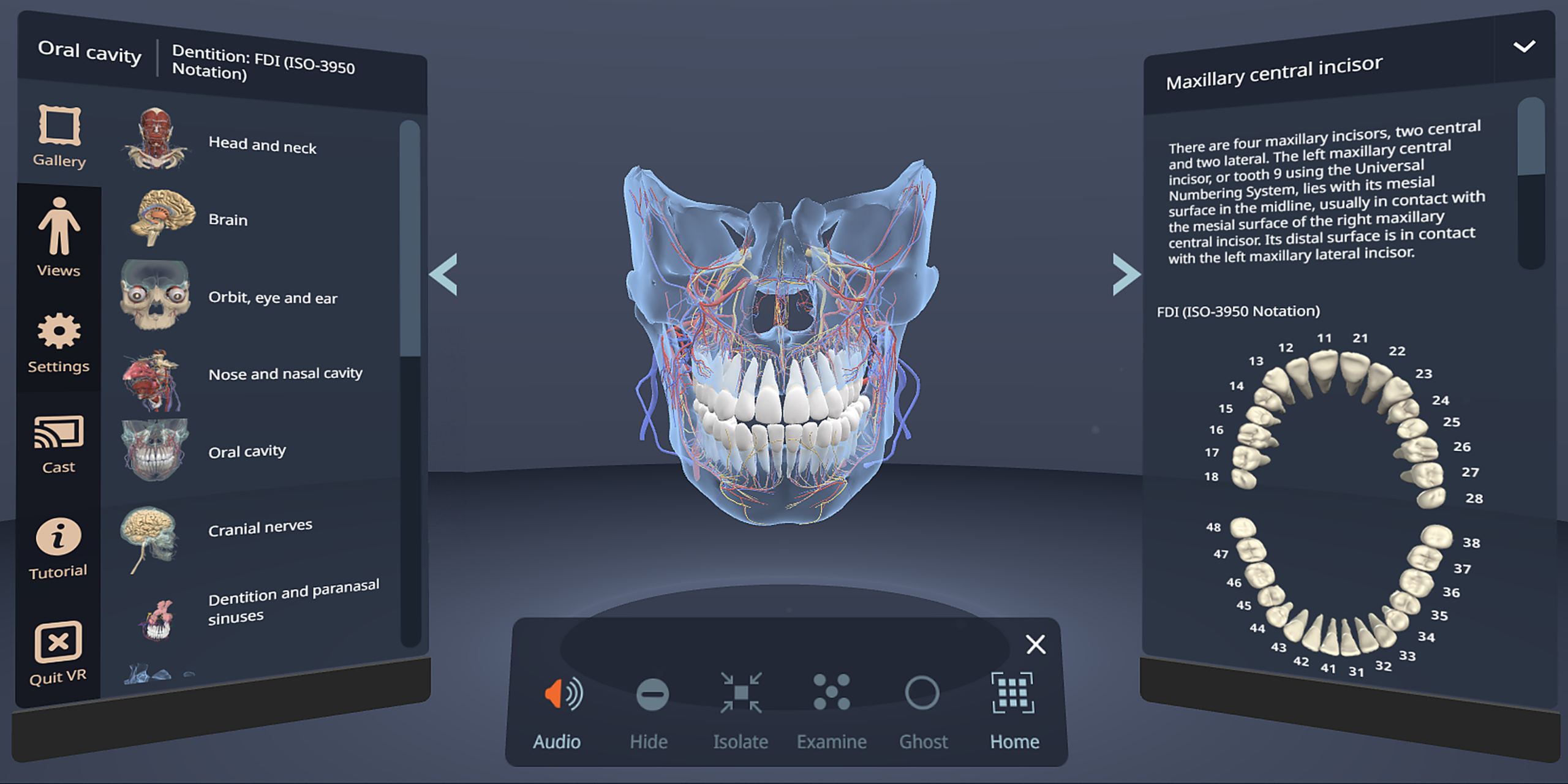
Gross anatomy teaching in dentistry is under real pressure. Access to dissection and prosection is declining across dental programs. Flat textbooks ask students to rebuild a three-dimensional oral cavity in their heads. And many will reach clinical placements without a reliable mental model of where structures sit.
The stakes are specific. Knowing which nerve to block, which structures sit in the path of an injection, and what can go wrong separates a safe procedure from a complication. That understanding must exist before a student touches a patient.
Primal Pictures addresses this through our dental portfolio. Our desktop content already includes fully interactive dissection images, so students can work with structures and read anatomist-written text. Now Dentistry VR closes the remaining gap by placing students inside the anatomy for true spatial understanding in an immersive environment.
Why is dental anatomy so hard to teach?
Three pressures make anatomy one of the hardest subjects to teach in dentistry:
- The oral cavity is small, confined, and spatially dense – Structures sit close together in three dimensions. Students without strong spatial reasoning have no reliable way to build an accurate mental map from 2D resources alone.
- Anatomy has to come before clinical skill – Dentistry asks students to find some structures and avoid others, sometimes in the same procedure. For example, to perform an inferior alveolar nerve block, the mandibular foramen must be identified by finding the medial ramus and using the pterygomandibular raphe as a soft tissue guide, while simultaneously avoiding the inferior alveolar vessels, the periosteum, and the parotid gland posteriorly. There is little room to develop that spatial model in parallel with practice.
- Access to hands-on alternatives is declining – Dissection and prosection access continues to narrow across dental programs, which removes the last physical route to spatial confidence.
The result is a gap between the anatomy dental students are taught and the anatomy they need to apply safely in the clinic. Closing that gap is where Dentistry VR shines.
What is Dentistry VR?
Dentistry VR is an immersive virtual reality learning application for teaching gross dental anatomy, built by Primal Pictures and delivered on Anatomy.tv. It runs in browsers on standard VR headsets. Students step inside true-scale 3D models of the oral cavity and the surrounding head and neck, and explore and interact with structures.
The module concentrates on the areas where spatial confidence matters most in dentistry: tooth morphology and the confined spaces of the head and neck.
Six preset Gallery Scenes cover the key head and neck regions, and a Views tab breaks each region into its component structures (e.g., a cranial nerves scene opens into a trigeminal nerve view).
Additional content includes:
- 3D tooth morphology gallery views
- A cross-section view of the tooth
- The dentition numbering system
Inside the experience, you can rotate, scale, and move models, and then isolate, ghost, or dissect individual structures. Show or hide arteries, veins, nerves, and lymphatics, or move through layers of anatomy one at a time.
Every structure also carries descriptive text written by anatomists and aligned with core anatomical textbooks, and includes an audio pronunciation guide.
Dentistry VR is designed for short, focused sessions of roughly 5 to 20 minutes. Use it for guided demonstrations, instructor-led exploration, small group teaching, or targeted self-directed study outside of class.
How does Dentistry VR work in education?
Give educators a way to show what they can otherwise only describe. For example:
- Guided gallery scenes and a ‘views’ tab present a clear path through the head and neck regions students find hardest, instead of leaving them to navigate a full model unaided.
- Isolate, ghost, and layer controls let you strip a dense region down to the structures that matter, which is hard to do clearly with static images.
- Short sessions of 5 to 20 minutes fit compressed teaching hours and are perfect for when students struggle to visualize structures or spatial relationships from traditional materials.
- Pass-through mode keeps VR practical in a normal teaching space, so programs do not need a controlled VR lab.
- Both guided and independent use are supported. Educators may run demonstrations and small group sessions, and students can return on their own for targeted study.
- Pairing with desktop 3D Real-time Dentistry on Anatomy.tv lets Dentistry VR build first-hand spatial understanding, while the desktop tools support reference, study, and clinical application. The two work together across different learning moments.
How is Dentistry VR different from other VR and 3D anatomy tools?
Dentistry VR sets itself apart from other platforms in a few specific ways:
- It places students inside the anatomy – They experience depth, scale, and spatial relationships directly, rather than reconstructing them from a flat textbook or a model on a desk
- A pass-through mode blends the model with the real room – Stay aware of your surroundings and see those around you, which keeps sessions more comfortable and practical in an ordinary classroom and helps limit any uncomfortable motion that some may experience. (Pass-through mode can be toggled on/off.)
- The content is clinically grounded and academically sourced – Every structure carries anatomist-written descriptive text and pronunciation audio.
- It is browser-based and device-agnostic – No software to install and no specific headset model requirements, which removes setup and hardware barriers that affect other VR products.
- It protects institutional hardware investment – Because the platform is not tied to one system or headset model, any program that changes headsets can still access the content.
Why VR when desktop 3D already exists?
Take the mandibular nerve, for example. It provides sensory fibers to the mandibular teeth, to the lower face, lower lip and temporal region, mucosa of the anterior two thirds of the tongue and floor of the mouth, and supplies the muscles of mastication. Textbooks describe its origin, path, and branches. 3D Real-time Dentistry lets you rotate, isolate, and remove everything around it to visualize it fully. Most students build a working mental picture from that.
But some do not. A student can name every branch of the mandibular nerve and still not know where it sits relative to regions of the mandible, the root above it, or the arteries beside it. That gap is spatial. More description will not help, and a better diagram won’t either, because both ask the student to perform the same mental reconstruction.
Dentistry VR removes the reconstruction step. At true scale, viewing from inside the body, depth and position are clear. The student sees where the nerve runs, what it runs between, and how much room there actually is.
Dentistry VR is not intended to replace our desktop tools, which include reference text, dissection images, and clinical content that the module does not. Dentistry VR complements by facilitating a spatial understanding when concepts resist visualization.
What does the research say about VR and 3D anatomy learning?
The case for immersive, 3D anatomy learning rests on a growing body of peer-reviewed research:
3D visualization improves learner satisfaction and engagement – Wang et al. (2024) ran a meta-analysis of randomized controlled trials on 3D visualization for human anatomy and found reliable gains in learner satisfaction and enjoyment over traditional 2D methods. Published in BMC Medical Education (DOI 10.1186/s12909-024-05403-4).
VR shows promise for anatomy learning outcomes – Odogwu et al. (2025) reviewed VR simulation against conventional anatomy teaching and found it a promising route to better outcomes — including spatial understanding, knowledge retention, and learner engagement — with 71% of the analyzed studies showed statistically significant learning gains using virtual reality. Published in Cureus (DOI 10.7759/cureus.81893).
‘Embodied cognition’ explains why physical involvement changes the learning – Embodied cognition means using bodily states and environmental interaction to support thinking, which is the theoretical foundation for why physically manipulating anatomy in VR produces better spatial understanding. Wilson (2002) concludes that several claims within the embodied cognition concept hold up under scrutiny: cognition is situated, it is time-pressured, and we offload cognitive work onto the environment. Applied to anatomy, this means a student thinks differently when standing inside a structure in VR than when reading about it. It also means manipulating a model in real-time, directly in VR, reduces the mental effort necessary for learning the relation of structures to each other. Published in Psychonomic Bulletin & Review (DOI 10.3758/BF03196322).
‘CAMIL’ explains why immersive VR benefits learning – Makransky and Petersen (2021) propose the Cognitive Affective Model of Immersive Learning (CAMIL) is also influential. The core argument is that VR activates specific cognitive and affective processes — presence (feeling “there”), agency (feeling in control), and embodiment (using your body to interact) — which in turn influence learning through increased interest, motivation, and deeper encoding. Published in Educational Psychology Review (DOI 10.1007/s10648-020-09586-2).
Who is Dentistry VR for?
Dentistry VR is built for four main groups:
- Dental programs under dissection pressure – Programs where dissection and prosection access is limited or declining, and educators need a credible resource that layers onto the hands-on instruction that remains.
- Educators teaching spatial anatomy – Instructors running demonstrations and small group sessions at the moments students struggle to visualize a structure.
- Students who need spatial confidence – Pre-clinical and clinical dental students building a mental model of the oral cavity before placements.
- Simulation, skills lab, and digital learning teams – The people who resource and support immersive teaching across a program.
Where does Dentistry VR fit within Primal’s entire dental portfolio?
Each dentistry module adds a layer of understanding, from foundational science to immersive spatial confidence.
| Stage | Content | Product |
|---|---|---|
| 1. Foundation | Core science base; how structures work and why | Human Anatomy & Physiology |
| 2. Guided 3D anatomy | Dental-specific anatomy with interactive layers, clinical text, and dissection images | Dentistry, Dental Hygiene |
| 3. Bespoke 3D anatomy | Freedom to rotate, ghost, and dissect structures; self-assessment | 3D Real-time Dentistry, Primal’s 3D Anatomy Quiz |
| 4. Immersive VR | A view from inside the anatomy for spatial confidence | Dentistry VR |
| 5. Clinical application | Treatment planning, patient communication, and procedure preparation | Dentistry, Dental Hygiene, Disease & Conditions |
Summary: What Dentistry VR delivers
| For Students | For Educators |
|---|---|
| Step inside true-scale gross dental anatomy | A credible resource that layers onto existing teaching |
| Spatial understanding that flat resources cannot build | Works in a normal classroom, with no dedicated VR lab |
| Dental-specific content covering tooth morphology, dentition, and the head and neck | Browser-based, with no installs |
| Anatomist-written text and pronunciation for every structure | Not locked to one headset, protecting hardware investment |
| Higher satisfaction and engagement than 2D methods (BMC Med Educ, 2024) | Pairs with additional Anatomy.tv desktop dentistry modules |
| Anatomy experienced, not just studied | Content trusted by 1,500+ institutions across 150+ countries |
Sources Cited
Makransky, G., & Petersen, G. B. (2021). The Cognitive Affective Model of Immersive Learning (CAMIL): A theoretical research-based model of learning in immersive virtual reality. Educational Psychology Review, 33(3), 937–958. https://doi.org/10.1007/s10648-020-09586-2
Odogwu, T. S., Abuelgassem Hagahmed Mohamed, E., Mishu, L., & Umahi, I. (2025). Effect of virtual reality simulation on anatomy learning outcomes: A systematic review. Cureus, 17(4), e81893. https://doi.org/10.7759/cureus.81893
Sinou, N., Sinou, N., & Filippou, D. (2023). Virtual reality and augmented reality in anatomy education during the COVID-19 pandemic. Cureus, 15(2), e35170. https://doi.org/10.7759/cureus.35170
Wang, J., Li, W., Dun, A., Zhong, N., & Ye, Z. (2024). 3D visualization technology for learning human anatomy among medical students and residents: A meta- and regression analysis. BMC Medical Education, 24, 461. https://doi.org/10.1186/s12909-024-05403-4
Wilson, M. (2002). Six views of embodied cognition. Psychonomic Bulletin & Review, 9(4), 625–636. https://doi.org/10.3758/BF03196322
Frequently Asked Questions
What is Dentistry VR? It is a browser-based virtual reality application on Anatomy.tv from Primal Pictures for teaching gross dental anatomy. Students explore true-scale 3D models of the oral cavity and surrounding head and neck structures, using natural movement to understand spatial relationships.
Who is Dentistry VR for? It is built for dental school programs and dentistry instructors, and for the pre-clinical and clinical dental students they teach.
What dental-specific content does it include? All relevant structures in the head and neck regions, 3D tooth morphology views, a cross-section view of the tooth, the dentition numbering system, and more. Every structure includes expertly-reviewed descriptive text written by an anatomist, and pronunciation audio.
What hardware do you need to run it? Most VR headsets are compatible. It runs in browsers, and does not require a dedicated VR lab, specialist PCs, or software installs.
Where does Dentistry VR sit alongside the other dental products? VR unlocks spatial understanding, and Primal’s additional desktop dentistry products reinforce and extend its content.
Does it replace desktop anatomy tools? No. It works alongside desktop tools such as Anatomy.tv’s Dentistry, Dental Hygiene, and 3D Real-time Dentistry, which support reference, revision, and clinical application.
Does it include assessment or clinical content? No. It focuses on gross dental anatomy and spatial understanding. Assessment is available through Primal’s 3D Anatomy Quiz, while clinical, procedural, and animated content lives in the desktop tools.
Is it for teaching or independent study? Both. It supports instructor-led demonstrations and guided group sessions, and students use it for short, targeted self-directed exploration.
How long are typical sessions? Short and focused, usually 5 to 20 minutes, used at key teaching moments rather than continuously.