What comes to mind when you think “appendix”? Perhaps you recall the story of a distant relative whose appendix “burst,” requiring emergency surgery to remove it. Maybe you fleetingly worry that the twinge of pain in your lower right abdomen could be a bout of appendicitis coming on. However little you may know about the appendix, you may have the idea that it is vestigial in nature. Like the wisdom tooth or tailbone, have humans evolved beyond needing its original function? Is its presence in the body merely a holdover from a bygone era?
Research conducted in the past couple of decades indicates that the appendix, also known as the “vermiform” appendix, still has an important role to play in the modern human body. To understand its possible vital contributions, let’s examine this often-misunderstood organ in greater detail.
The content in this post is from Primal’s 3D Atlas module. To learn more about this or other Primal learning resources, please fill in the form here and our team will be in touch.
What is the anatomy of the vermiform appendix?
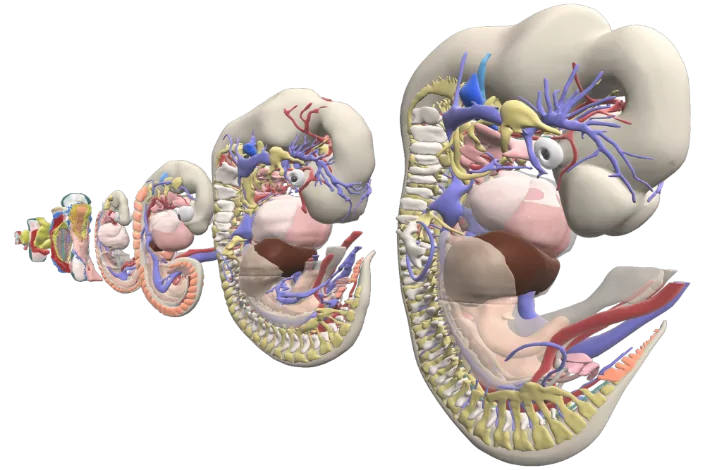
The appendix typically makes its first histological appearance in the human body around the eighth week of gestation. It then undergoes several rotations and adopts various positions along with the developmental elongation of the rest of the large intestine.
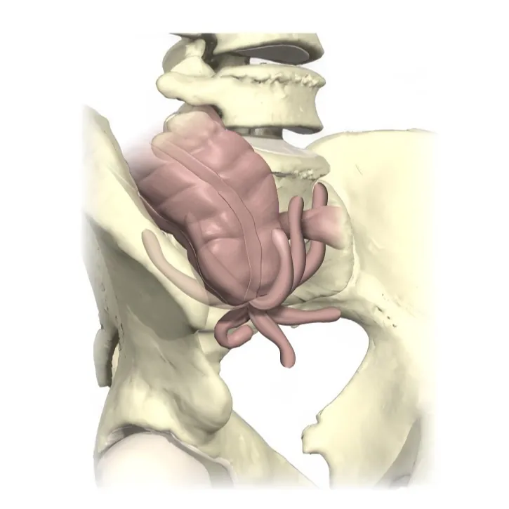
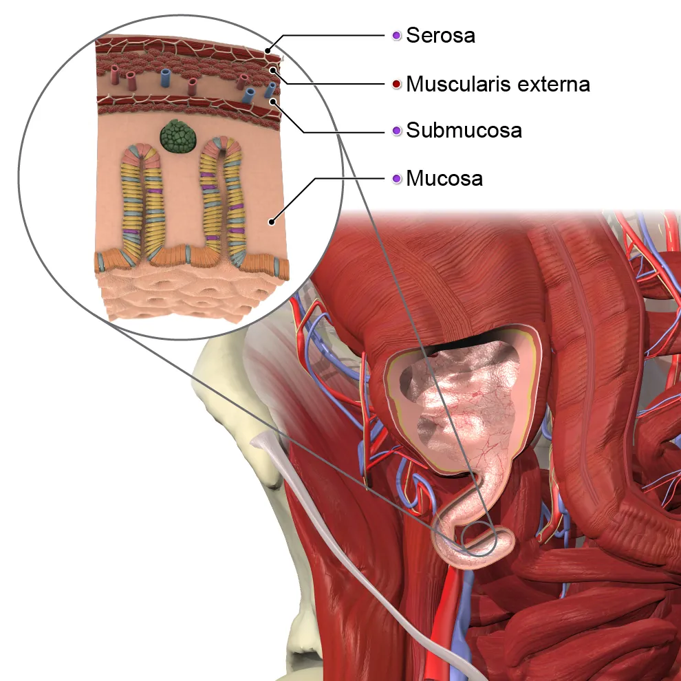
Usually located in the lower right corner of the abdomen, the vermiform appendix is a tube of soft lymphoid tissue with one end closed (making it more of a pouch). It consists of the base , body, and apex (tip), and is comprised of four layers:
- Mucosa (inner layer)
- Submucosa (connective tissue beneath the mucosa)
- Muscularis externa (smooth muscle)
- Serosa (outer layer)
Its open end leads into the cecum, or first part of the large intestine. During digestion, the cecum is the section of the large intestine into which the small intestine empties its contents. Although variations can exist, the vermiform appendix is approximately 9 cm, or 3.5 inches in length, making it roughly the same size and shape as a finger. If you use your imagination, it resembles a small snake or worm. In fact, “vermiform” is Latin for “worm-like.”
What variations of the appendix are found in the body?
While the location, size, and shape of the appendix described above is most commonly seen, researchers have found a number of variations in the presentation of this somewhat mysterious organ. Since it is only attached at one end, its positioning in the body may differ:
- Retro-cecal – the tip of the appendix appears behind the cecum (the most common variation, found in approximately 44% of people)
- Pelvic – the tip of the appendix points downwards into the pelvis
- Sub-cecal – the tip of the appendix is just below the cecum
- Para-cecal – the appendix is positioned next to the cecum
- Pre-cecal – the appendix is positioned in front of the cecum
- Pre-ileal – the appendix is positioned in front of the ileum
- Post-ileal – the appendix is positioned behind the ileum
- Sub-ileal – the appendix is positioned under the ileum

In very rare cases called situs inversus, the appendix may even be positioned on the left side of the abdomen rather than the right.
Along with variations in location, appendixes can also vary in size and shape, likely influenced by factors such as genetics, geography, sex, diet, and race. In some cases, an individual may be born with an underdeveloped appendix. Others may be born without an appendix at all.
What does the appendix do?
Although the presence of the appendix has been recorded as far back at the late 15th century, only recently have scientists begun to think of the organ as more than a vestige of the past. Until about 20 years ago, Charles Darwin’s theory about the appendix prevailed. He thought it was part of a previously much larger cecum, once necessary for when ancient humans subsisted on mainly a diet of leaves, but rendered obsolete when early human diets became more fruit-based.
But more recent studies have posited that the development of the appendix, home to an abundance of gut flora, may have occurred prior to the development of the cecum. What does this mean? Scientists now believe that the appendix may have initially played — and continues to play — a key role in the body’s immune system, rather than being a mere remnant of the digestive system. Research has shown that the appendix helps stimulate the development of gut-associated lymphoid tissue (GALT). GALT is critical to helping the gut’s immune system monitor and respond to bacteria, detecting and fighting against harmful substances while identifying and helping the body tolerate harmless or helpful bacteria in the gut. This begins a few weeks after birth, when bacterial translocation begins and bacteria moves from the intestines through the intestinal mucosa to the mesenteric lymph nodes. This introduction of bacteria from one area to another is thought to be an essential element in training immune cells to learn which microbes are helpful and which are harmful. Alongside the appendix’ involvement in development of GALT, it is also found that biofilm, a layer of commensal gut bacteria believed to help the immune system exclude pathogens. This biofilm also acts as a reservoir for harmless or even beneficial bacteria, which is deployed to “rebalance” the system after intestinal illnesses.
What clinical complications are associated with the appendix?
While the appendix may contribute to the strength of the intestinal immune system, because of its coiled structure and the presence of bacteria within, it can also be a hotbed of infection. Inflammation of the appendix is called appendicitis. Appendicitis is a global disease, with evidence correlating its rise in newly industrialized societies throughout the world with the environmental factors typically present in those areas.
Acute appendicitis occurs when the hollow space inside the appendix becomes blocked, usually by an overabundance of cells (lymphatic hyperplasia) that occurs in reaction to inflammatory conditions like a virus. In rare cases, a parasitic worm infestation, such as enterobius vermicularis, can also trigger acute appendicitis. Older people may also display symptoms similar to those of acute appendicitis when arteriosclerosis, which results in build-up of fatty plaques in the arteries, causes hardening and decreased blood flow to the appendicular artery. This can deprive the nutrient and oxygen supply of the appendix, damaging its cells and triggering an acute inflammation reaction. Typical symptoms of acute appendicitis include loss of appetite and nausea, followed by intensive pain in the lower right abdomen (for patients whose appendix is in the usual spot).
Treatment for acute appendicitis is usually the immediate surgical removal of the appendix — which can be tricky to find, given the variations in its location, size, and shape as discussed earlier. It is vital that surgeons be aware of these variations so they can accurately and quickly locate and remove the inflamed appendix. This is especially crucial as an inflamed appendix can rupture and spread the infection across the abdomen, a condition called peritonitis. This life-threatening condition can happen quickly, as soon as 48 to 72 hours after you first notice symptoms. So remember: if you suspect that pain in your side is appendicitis, don’t hesitate to take action.
The content in this post is from Primal’s 3D Atlas module. To learn more about this or other Primal learning resources, please fill in the form here and our team will be in touch.