

The act of hugging — whether from a friend, relative, or romantic partner — can be a simple expression of affection, protection, or comfort. But the anatomy and physiology of this gesture is surprisingly complex. Let’s take a deeper look.
What is involved in the act of hugging?
A parent comforts a crying toddler. Old friends greet each other after a long absence. Partners settle in for the night. While each of these embraces may happen spontaneously and organically, the mechanisms at play during a hug are actually quite complex. Far from merely the extension of the arms and contraction of a few muscles, a hug is instead a highly orchestrated physical action involving not only muscles and nerves, but also the release of neurochemicals and hormones. To warmly embrace someone, the body must coordinate movement while gathering and transmitting information to and from the nerves and the brain — all while stimulating the emotions. The three aspects of movement, touch sensing, and chemical responses make up the core components of a hug.
What happens first in a hug?
Before we can enjoy the feelings of love, calm, or peacefulness that are often triggered by an embrace, our bodies must first be set into motion. Hugging requires the harmonized actions of muscles across several regions, mainly the arms, hands, and shoulder girdle (the scapulae/shoulder blades and clavicles). During this coordinated action, the shoulders move forward while the arms wrap inward and the hands adjust their grip.
In preparation for a hug, the arms move away from the body at the upper arm’s glenohumeral joint in a motion known as abduction. Meanwhile the scapulae begin to slide outward around the rib cage, driven primarily by the serratus anterior. As the arms start to sweep forward and inward, additional muscles contribute to drawing someone close. The latissimus dorsi, a broad back muscle that assists with arm adduction and internal rotation, helps generate the inward pull that brings the person toward the body.
At the same time, although the scapulae protract to wrap the arms around someone, they must remain stable against the ribcage. This stability is provided by deeper muscles such as the rhomboids and portions of the trapezius, which work together to maintain proper scapular position and prevent the shoulder blades from winging or drifting excessively during the embrace.
After the arms open, they then begin to move forward, and the motion changes from abduction to horizontal adduction, pulling the arms back to the body’s midline. Next, the arms move inward, pulled forward by the pectoralis major muscle and assisted by the anterior fibers of the deltoid muscle. As the person being hugged gets pulled closer, the elbows flex, powered mainly by the biceps brachii muscles. Finally, the flexor digitorum superficialis and profundus muscles work to flex the fingers, creating tighter grip on the person being hugged.

How is the sense of touch processed during a hug?
The act of hugging encompasses more than just simple physical mechanics; hugs often elicit powerful emotional reactions as well, thanks to nerve receptors on the skin that act as an input system to the brain.
These cutaneous receptors respond differently to different types of sensory input, helping to affect emotions in various ways as they relay information to the brain. The type of cutaneous receptor that responds to touch or pressure is called a mechanoreceptor.
Mechanosensory neurons transform mechanical forces on the skin into electrical signals that are then sent to the brain, with different types of mechanoreceptors responding differently depending on the type of touch detected. The types of mechanoreceptors involved in hugging include:
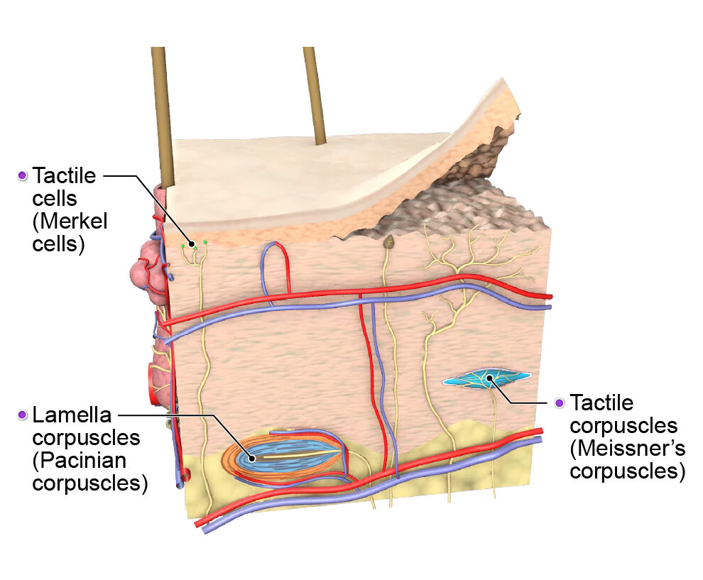
- Tactile corpuscles (Meissner’s corpuscles) – These dendrite masses are found in their greatest numbers on hairless skin, such as the hands and inner arms. They respond to light touch and movement.
- Tactile cells (Merkel cells) – These cells are associated with a sensory nerve ending called a Merkel disc (or tactile disc) and respond to sustained pressure (holding).
- Lamella corpuscles (Pacinian corpuscles) – These cells rapidly adapt to stimuli and respond to deeper pressure and vibration. They are found in the subcutaneous layer of the skin and in deeper regions, such as the muscles.
Another, distinct type of cutaneous receptor responsible for sending signals to the brain during a hug is a C-tactile (CT) afferent. These represent a separate, specialized pathway, transmitting pleasure signals toward the brain, letting us know that a light caress or gentle hug feels good. They are found in skin with hair and are most sensitive to slow, gentle, skin-temperature contact.
What happens to these signals after they are transmitted to the brain?
The physiological effects of a hug are not instantaneous. Signals of touch are first transmitted by the cutaneous receptors via the spinal cord to the brain’s insula, located deep within the lateral sulcus. The insula is involved in interoception (sensing, interpreting, and responding to internal bodily signals — the “inner sense”), affective touch, and emotional processing. It is responsible in part for visceral sensation, and it is in the insula that the brain processes not only the awareness of the body being touched, but also the emotional reaction to that sensation.
During a welcome embrace, the insula processes signals trigger a variety of stress coping mechanisms to help calm the body. These include lowering blood pressure, reducing the heart rate, and controlling certain hormone levels, all of which contribute to an overall feeling of peacefulness and positivity. Essentially, a welcome, warm embrace lowers the response of the body’s sympathetic nervous system (responsible for “fight-or-flight” reactions such the production of adrenaline, rapid breathing, and being in a state of high alert or nervousness). At the same time, the tactile stimulation of a hug increases the activity of the parasympathetic nervous system, reducing signs of anxiety.
This parasympathetic activity includes stimulating the release of oxytocin, sometimes called the “love hormone.” Oxytocin is an anti-inflammatory neuropeptide molecule that is produced in magnocellular neurons of the hypothalamus and released through the posterior pituitary gland at the base of the brain. In pregnant people, it is instrumental in triggering labor contractions and aids in milk release during breastfeeding. But the calming effects of oxytocin are not limited to those giving birth. It also gets released in all people as a response to nurturing, affectionate, or sexual moments of connection — like an embrace — flooding the body with feelings of wellbeing. Positive touch such as hugging has also been shown to reduce the body’s levels of cortisol. Cortisol is the body’s main stress hormone, which is produced by the adrenal glands. It helps the body regulate blood pressure and glucose levels and supports the body’s recovery after a stressful event has passed. A hug sends signals of safety to the brain, reducing the need for cortisol and communicating that it is safe to move out of a state of stress.
When should we ‘hug it out’?
While research shows that hugging can increase feelings of calm and reduce signs of stress, not everyone may be down for “bringing it in.” Although embraces among friends, acquaintances, or family may be prevalent across some cultures and are an important means of social interaction, hugging remains a rarity — even among close family members — in others. Some people may prefer not to hug because they have sensory sensitivities that make certain feelings of touch uncomfortable. Others may have a difficult time returning a hug due to pain, limited range of motion, or injury that restricts movement or makes certain movements hurt. If there is any doubt of how your well-intended hug may be received, asking first can avoid possible embarrassment or discomfort. And if you wish to incorporate more hugging into your life, but find that doing so is difficult for you physically, have a chat with your doctor to see if physical therapy could help.