
As temperatures rise and spring arrives in many parts of the world, breathing in a bit of fresh air after a long winter is a welcome ritual. But what exactly happens within the lungs as we take a deep breath, and what can advanced imaging show us about this vital process and the essential structures which support it?
How does the air that we breathe impact the lungs?
While taking a deep breath may feel rejuvenating, the quality of the air we breathe is crucial to the health of our lungs — a fact highlighted each year on April 22, Earth Day. Environmental hazards like air pollution can negatively impact the structure and function of our respiratory system. These environmental impacts are difficult to escape: the World Health Organization (WHO) estimates that 99% of the global population breathes in air that exceeds the limits of WHO safety guidelines. Exposure to particulate matter air pollution is responsible for an estimated 9 million premature deaths per year, exceeding the number caused by diseases like high blood pressure or diabetes. Depending on the severity of the impact, the lungs and airways can have temporary or chronic damage. Therefore, detecting changes in lung structure is essential to accurately diagnosing issues within the lungs as quickly as possible. Diagnostics like X-rays and CT scans play a critical role in accurately identifying these changes in time.
What is the anatomy of the respiratory system?
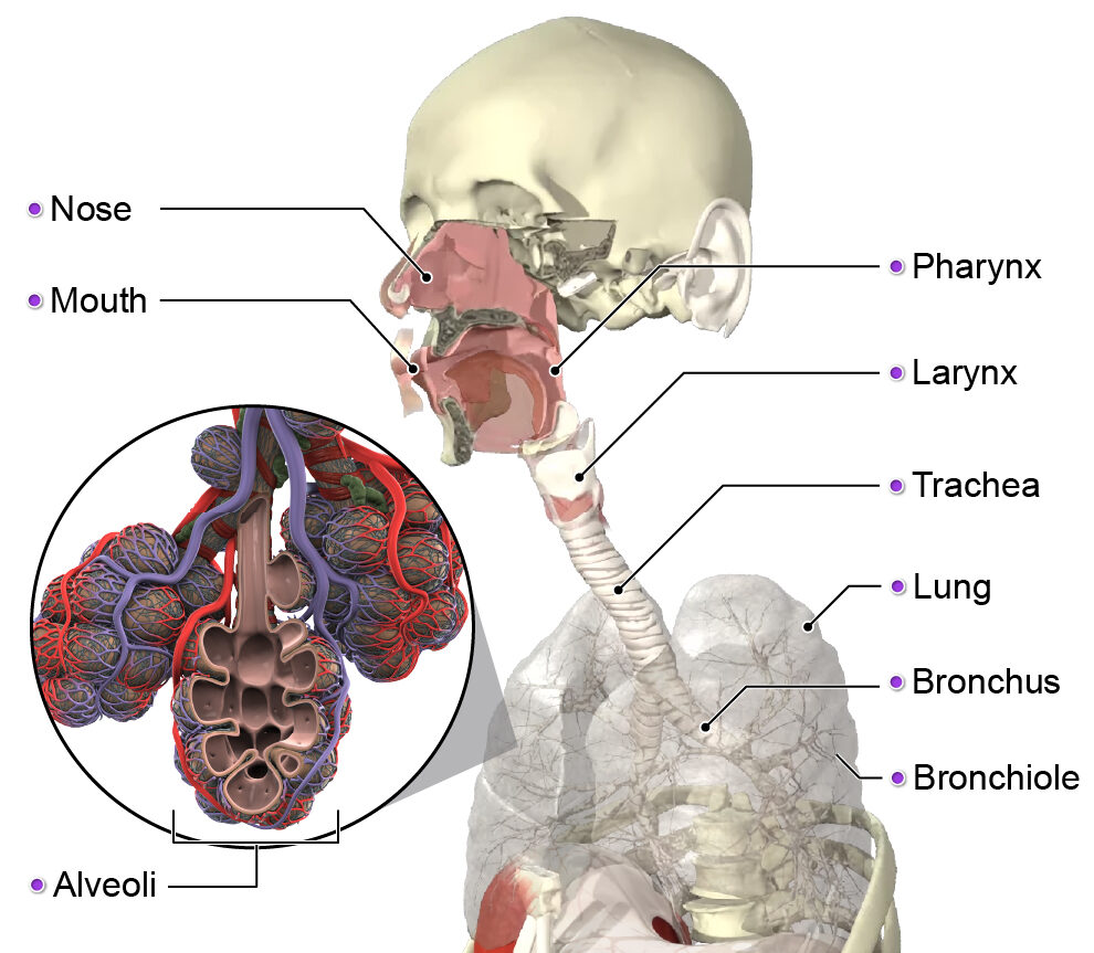
When we breathe in (inhale), air must pass through several structures before eventually reaching the lungs. The air enters our body via the upper respiratory tract, which includes the mouth, nose, and pharynx.
After air is taken in through the upper respiratory tract, it passes through the structures of the lower respiratory tract, including:
- Larynx: A cartilaginous structure often known as the “voice box” due to its primary role in supporting voice generation. It also has several important functions related to breathing, including protecting the airway and closing and sealing the lower respiratory tract.
- Bronchial tree: Originates directly beneath the larynx. It is comprised of the trachea, or windpipe, which divides into bronchi. The bronchi then divide further into bronchioles, which control air flow and the distribution of air in the lungs. Each bronchiole has several alveoli, which are thin-walled air sacs that are rich in blood supply. Alveoli are the main site for gas exchange, which is a process of exchanging oxygen and carbon dioxide. Human lungs contain over 300 million alveoli.
The air then finally encounters the lungs, which consist of two sets of specialized spongy tissue, and is located within and on either side of the mediastinum — the middle section of the thoracic cavity (chest). The lungs rest on the diaphragm and are protected by the ribs.
Although both the right and left lungs perform the same tasks, they differ in their shape and structure to accommodate other nearby vital organs like the heart and liver. Main structures of the lungs include:
- Lobes: The right lung has three lobes (superior, middle, inferior), while the left has two (superior, inferior).
- Fissures: These are the narrow depressions that divide the lungs into lobes. The right lung has two while the left lung has one.

How do you view lung function?
Using imaging tools is critically important to detecting and diagnosing diseases or malfunctions of the lungs. Typically, clinicians rely on chest X-rays (or chest radiography), computed tomography (CT) scans, or to a lesser extent, magnetic resonance imaging (MRI) scans to gain a view of lung function or abnormalities.
Standard chest X-rays will show certain key visible structures of and around the lungs, including features like the trachea, the diaphragm, and the lungs themselves. Chest X-rays are also notable for what they do not show: While an X-ray of a healthy chest will not show structures like the pleura or aorta, these structures will become visible on the X-rays of unhealthy lungs, indicating the presence of possible disease or other conditions negatively impacting lung function.
While clinicians commonly use chest X-rays as the initial imaging solution when a problem with the lungs is suspected, CT scans can provide a more detailed view of the lungs, creating two- or three-dimensional images that clinicians can use to detect possible issues. On CT scans, interstitial lung abnormalities (ILAs) appear as hazy areas of increased opacity, called ground glass opacities. Depending on their pattern, they may indicate certain lung diseases. CT scans are also useful in showing more detailed images of unusually sized airways. Non-invasive, fast, and widely available, thoracic CT images can provide a clear clinical view of the presence or absence of various lung diseases, tumors, pneumonia, and more.
When a closer look at the tissues of the lungs is necessary, clinicians will sometimes turn to MRI, which provides useful clinical data for soft tissues in certain situations. However, MRI technology has some significant limitations when performing lung scans. The lungs contain a large amount of air and a relatively small amount of tissue, making MRI — which relies on detecting water in tissue matter in order to create images — more difficult. Patient movement as well as the beating of the heart can also cause difficulties in capturing a clear lung MRI scan, although recent developments related to MRI pulse sequences may soon change that.
How do you monitor lung issues?
No matter which modality is used to obtain them, lung images provide clinicians with the essential information needed to not only detect irregularities, but also to diagnose diseases and to monitor their progression.
For instance, a diagnosis of pneumonia, a common lung ailment in which the lung tissue becomes inflamed and the alveoli get filled with fluid, is usually confirmed with a chest X-ray. The X-ray will show patchy shadows on the lungs where inflamed tissue has consolidated, preventing x-rays from passing through. Likewise, chronic lung diseases like chronic obstructive pulmonary disease (COPD) can be discovered and monitored using CT and MRI radiological markers. Certain markers like airway thickness parameters or perfusion defects (areas of the lungs which show a lack of blood flow) are useful indicators and measures of structural unusualness. Clinicians can use these to detect and quantify the presence of COPD, while at the same time ruling out other illnesses. These imaging insights can also be used to inform future clinical trials by giving researchers key information about the disease’s development and physiological effects on the body.
How does air quality affect lung health?
Beyond genetic predisposition or lifestyle choices like smoking or vaping that may increase the likelihood of lung disease or damage, air pollution has emerged as a pervasive risk factor for nearly all of the world’s population. Air pollution can be found both outdoors (ambient air pollution) and indoors. Ambient air pollution takes the form of gaseous components and airborne particulates that arise from activities or events like the combustion of fossil fuels, factory emissions, or wildfires. Indoor air pollution puts people at risk for lung damage via exposure to things like fireplaces and wood stoves, cooking activities, second-hand smoke, mold and bacteria, solvents and cleaning agents, and building materials themselves among many other sources.
The impact of air pollution on the lungs depends on both the nature of the pollution and the length of exposure to it. Shorter exposure to higher levels of particulate pollution can cause as much damage as long-term exposure to lower levels of pollution. Particulate matter pollution is most harmful to the lungs when the particles are smaller in size and therefore able to more easily penetrate bronchioles and alveoli. This can cause damage to the alveoli walls, inflammation of lung tissue, oxidative damage to the lung tissue, dysregulation of the immune response, and even alterations to DNA. Researchers have linked the damage from both short- and long-term exposure to air pollution to increased incidence of lung cancer, pulmonary fibrosis, chronic respiratory diseases like COPD and asthma, and other conditions.
Earth Day is a timely reminder that lung health and the environment are inextricably linked. By taking steps today to limit the production of harmful airborne particulates and your exposure to them, you can ensure that you will breathe easier tomorrow.