Embryology is integral to medicine, dentistry, nursing, and other health sciences. But available learning resources – notably 2D textbooks – lack translation of the complex 3D morphing, twisting, and development of structures in the embryo. That’s where Primal’s new 3D Real-time Embryology can help.
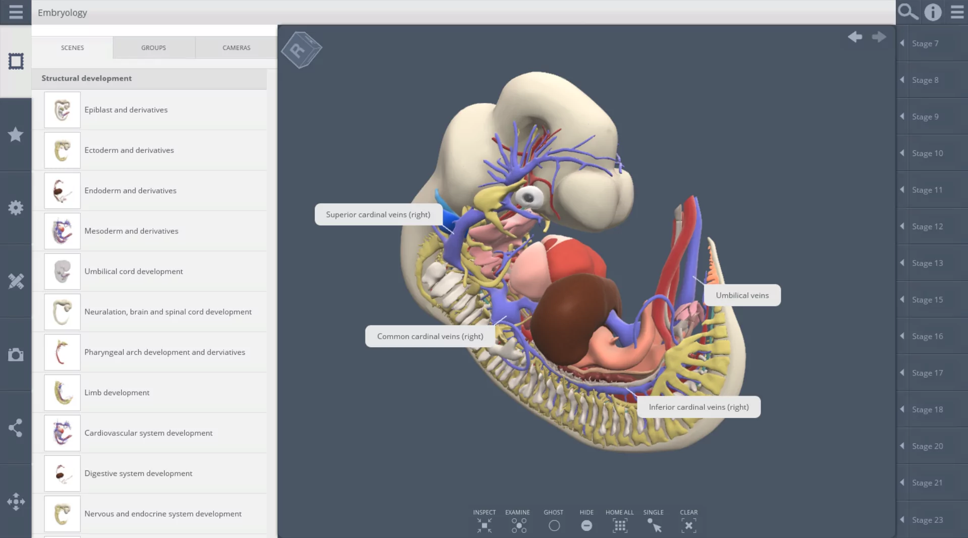
3D Real-time Embryology showcases these vibrant, 3D structural changes with real-scan data and corresponding terms that clearly define developmental changes at different stages to open up entirely new learning opportunities. Explore detailed 3D embryos from weeks 3 to 8 of development, when key organs and structures form.
Our 3D embryo models are sourced from Amsterdam University Medical Center (UMC)’s real-life data, which includes the Carnegie collection of over 10,000 specimens, images, and models. We’ve developed the models further with our in-house anatomy team and graphic designers to ensure they are accurate and visually engaging. The 3D structures can be dissected, examined, and labelled in our user interface.