
Anatomical education is evolving from static 2D diagrams to immersive, interactive experiences. Lauren Walsh, during her postgraduate studies in Medical Visualisation and Human Anatomy at the University of Glasgow, has explored how virtual reality (VR) and Primal VR can revolutionize the understanding of complex anatomical regions — specifically the orbit around the eye.
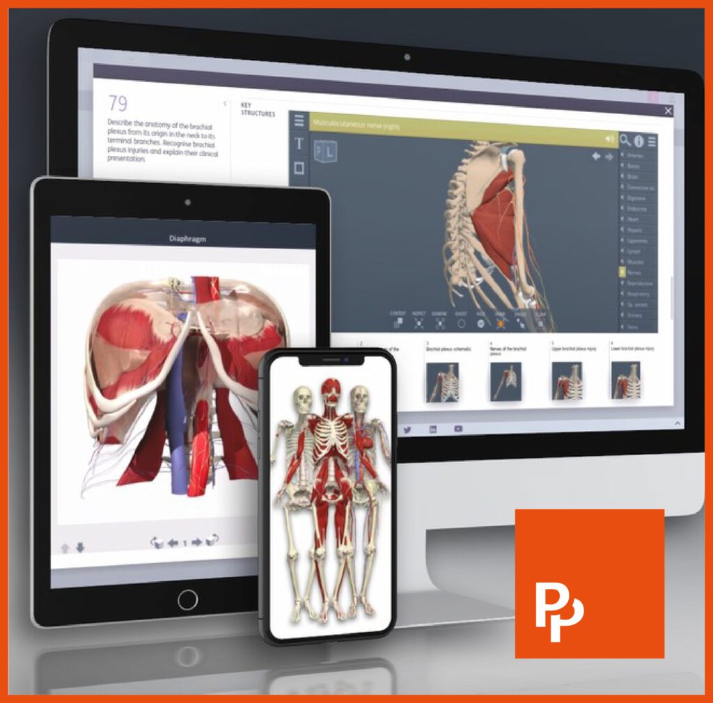
Her project, “Visual Pathways VR: An Immersive Exploration of the Orbit and Associated Traumas,” leveraged Primal’s highly detailed 3D anatomical models from Anatomy.tv. With Primal VR, she created an app that allows users to highlight, isolate, and animate anatomical structures, while referencing real dissection images.
For students working with tightly layered or clinically sensitive regions, VR can provide clarity that is difficult to achieve from diagrams and images alone. Integrating VR alongside dissection photographs or clinical imaging can further strengthen understanding.
Lauren Walsh
The result? Improved clarity and confidence in anatomy understanding, overwhelmingly positive feedback from colleagues, a scalable learning solution, and more. Learn more by reading the full case study, or contact us today for a demo or free trial.
This collaboration demonstrates how Primal’s 3D models, when combined with VR, can transform anatomical education and make it more engaging, intuitive, and accessible. It highlights the growing potential of XR-based tools for medical training and clinical skill development.