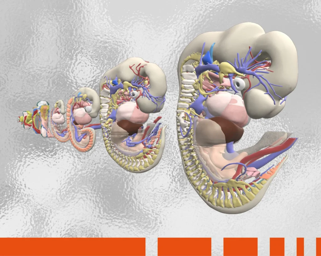
The medical world has come a long way from the crude skeleton models used as early as the 14th century. Today, such rudimentary skeletons are relegated to Halloween lawn and retail displays. Replacing these relics is 3D anatomy software that provides a far more accurate and realistic representation of the human form – beyond the bare bones.
Primal Pictures’ 3D anatomy model, built using real scan data from the Visible Human Project, has been carefully segmented to create an unparalleled level of detail and accuracy. All of the content within this program has been verified by qualified anatomists and by a team of external experts for each body area.
An obvious use for anatomically accurate software is academia. The study of human anatomy is not limited to those pursuing an M.D.; it is a core component of health science programs such as nursing, human kinetics, physiotherapy and occupational therapy. It is crucial for these students to fully understand the architecture and intricacies of the human anatomy.
To supplement classroom lectures, the traditional anatomical hands-on teaching method for the health sciences has been lab work — or, more specifically, the “C” word — cadaver labs. According to a University of Ottawa survey, however, this traditional approach is met with a myriad of challenges:
• Anatomy lab sessions require significant investments of time, space and resources
• These lab sessions are of limited duration
• A student’s view and/or ability to manipulate the cadaver is often restricted
• Due to large student enrollments and/or information-dense curricula, it is difficult to offer weekly anatomy labs
The survey authors conclude that “interactive learning tools can be tailored to meet program-specific learning objectives as a cost-effective means of facilitating the study of human anatomy. Virtual interactive anatomy exercises provide learning opportunities for students outside the lecture room that are of especial value to visual and kinesthetic learners.” For online learning, thanks to 3D anatomy tools, “zoom” takes on a new meaning, providing close-up views of the human body unlike any other. These tools also properly depict scale, literally giving students a brand-new perspective.
Consider this: The human body is made up of approximately 78 organs, 600 muscles, 900 ligaments, 4,000 tendons, over 200 bones, 100,000 miles of blood vessels and billions of nerves. Primal Pictures’ 3D Atlas captures it all, with unprecedented precision, in over 200 movies and animations, 435 views, 700 MRI images and 900 library assets. And while many traditional anatomical models show the healthy human body, Primal Pictures’ Disease & Conditions pathology module features 88 clinical conditions across multiple specialties.
A paper published in The Anatomical Record notes that: “Teaching anatomy by dissection is under considerable pressure to evolve and/or even be eliminated.” Taking its place are online interactive anatomy programs that “enhance the dissection experience, observational learning, and three-dimensional comprehension of human anatomy.” Like textbooks and traditional models, in lab specimens one structure must be removed in order to show another.
Virtual labs are now becoming de rigueur in anatomy curricula. A paper on “The Effect of 3D Human Anatomy Software on Online Students’ Academic Performance,” published in the Journal of Occupational Therapy Education, compared grad students’ performance in a traditional lecture/lab format (control group) vs. online learning (intervention group) plus four in-class lectures. The results? Final course grades were better for the intervention group as compared to the control group, regardless of age and learning style.
For those willing to think outside the lab, Primal Pictures’ 3D Real-time focuses on digital and cadaveric dissection. This interactive digital resource covers all anatomical regions with detail, accuracy and flexibility.
The concept of virtual reality (VR) has quickly moved from the gaming console to the classroom and many other applications. As an educational tool, VR enables students to learn through practical experience, which has been found to substantially increase the quality of retention and recall according to a University of Maryland study. VR bypasses the brain processes that distract the learner from true problem-solving and creative thought.
With VR, anatomy students can view structures from the inside out, adding and removing layers. VR allows for independent exploration, reinforcement and mastery of structures and systems. Primal VR, for example, is a fully immersive learning experience, combining Primal Pictures’ best-in-class 3D human anatomy model with cutting-edge VR technology.
Everyone absorbs information differently; some people grasp more by hearing, some by seeing (both reading and visuals), and others by doing. When healthcare practitioners need to explain complicated diagnoses and treatment plans to patients, it is important that they present the information in both a manner and format that are easily understandable. This is particularly true when breaking bad news; the patient will likely be in a distressed state of mind and need help with focusing on the options being presented.
They say a picture is worth a thousand words. When it comes to educating patients, 3D human anatomy images and 360-degree animations are worth multiples of thousands of words (most of which are “medicalese” gibberish to patients). Interactive visuals promote clear communication, resulting in improved patient engagement, trust, compliance and, ultimately, outcomes.

Chiropractor Ian Reed says, “It’s easy to forget that our training has given us a very detailed knowledge of how the body operates.” He uses Primal Pictures’ 3D human anatomy software to “bring the body to life” for patients.
“It’s a bit like leading the patient by the hand,” he says, adding that the images clarify patients’ understanding. A patient can relate the images to a point on their own body and can therefore make more sense of what is happening to them.
Healthcare companies whose customers include practitioners, hospitals, medical schools or training institutions must visually — and powerfully — demonstrate the value of their products and services. Whether the business focus is research and development, corporate training, product management, marketing, sales, end-user training, development or support, seeing is believing for customers.
Detailed human anatomy images, video and 360-degree animations can be used for a variety of training materials, including workbooks and manuals, classroom slides, video presentations and e-learning modules. A firm understanding of anatomy and physiology enables healthcare and life sciences product managers to effectively communicate product requirements, functionality, features and benefits to customers as well as to other team members and coworkers in other divisions.

From a marketing standpoint, engaging anatomy images and customized animations and videos can be reproduced clearly in presentations, sales demos and proposals; in digital content for social media, websites and video; and in printed materials such as brochures and product sheets. These assets also can be used to create compelling, branded, hands-on external training tools such as interactive videos, drill-down exercises, rotatable 3D animations and diagrams with embedded anatomy content.
Whether software for human anatomy is used to teach students, inform patients or impress customers, it adds a level of interactivity and engagement that cannot be equaled by one-dimensional print textbooks, crude plastic models or uninspiring slide decks. The coronavirus pandemic forever changed the way we communicate with one another; remote, online platforms are here to stay. It’s up to us, however, to make the most of these new mediums, turning challenges into opportunities. By embracing and harnessing technology, we’ll be able to envision a brighter future, one with better healthcare for all.