Clearly, not every institution is able to offer hands-on cadaveric resources to students, while those that are, need to ensure that the experience delivers full educational value. So, we’ve put together this essential guide to making the most of our digital dissection resources whatever the teaching environment.
This includes creating flexible and repeatable learning tasks that enhance and reinforce knowledge both inside and outside the lab. We’ve also got top tips on helping students prepare for, and revise after, in-person or virtual dissection sessions. Plus, we’ve included handy ways to create and conduct informal and formal assessments of what students have learnt.
Challenges Anatomy Educators Face Right Now
Most anatomy educators recognize that by getting up close to physical specimens, students gain practical skills and multidimensional understanding of the human body and anatomical variation, while learning aspects of professionalism.
However, some institutions do not have ready access to a working cadaver lab. Others have access but are currently unable to operate at full capacity owing to restrictions around COVID-19. What’s more, global pandemic or no global pandemic, the amount of time and number of specimens available to students in a physical dissection lab is usually limited.
All of which comes back to the same underlying challenge: ensuring that students have access to accurate, targeted, quality dissection materials in lab and remote settings, such as virtual seminars or while studying at home.
How Primal Boosts Spatial Awareness and More
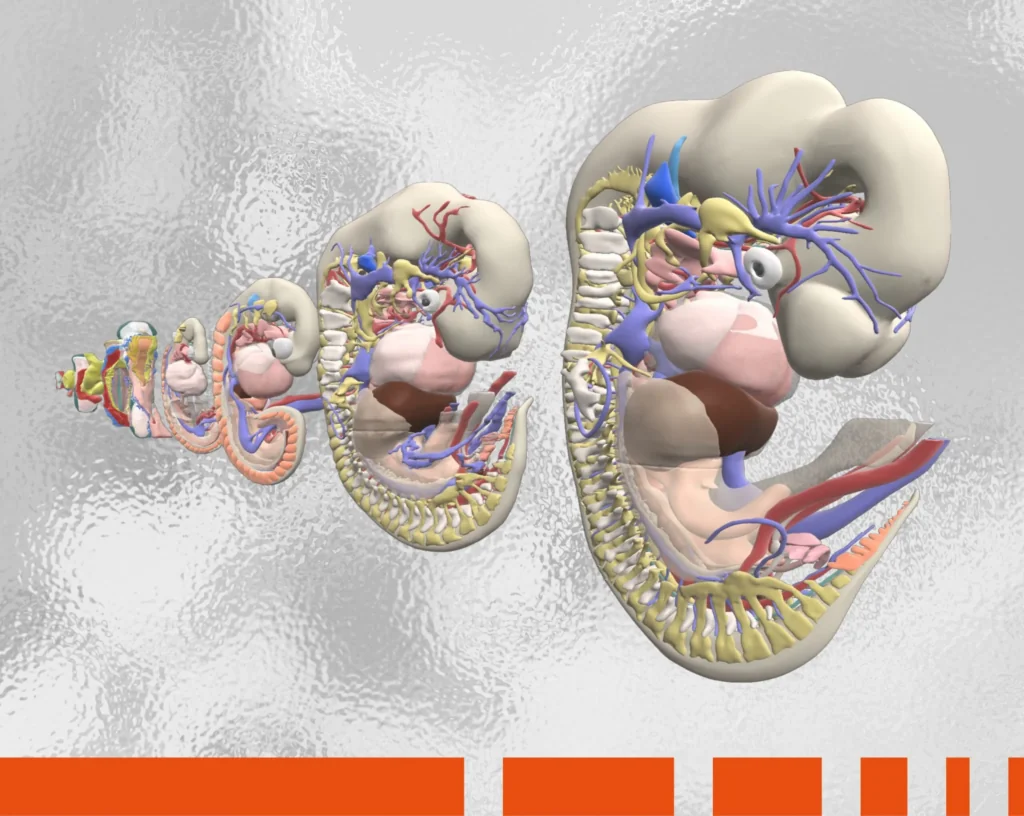
One of the main aims of using dissection resources with students is to develop their spatial awareness. Primal has a wealth of interactive and fully labeled digital dissection images that are specifically targeted at enhancing spatial ability. Indeed, they are designed to deliver beyond what even physical dissection of a cadaver achieves.
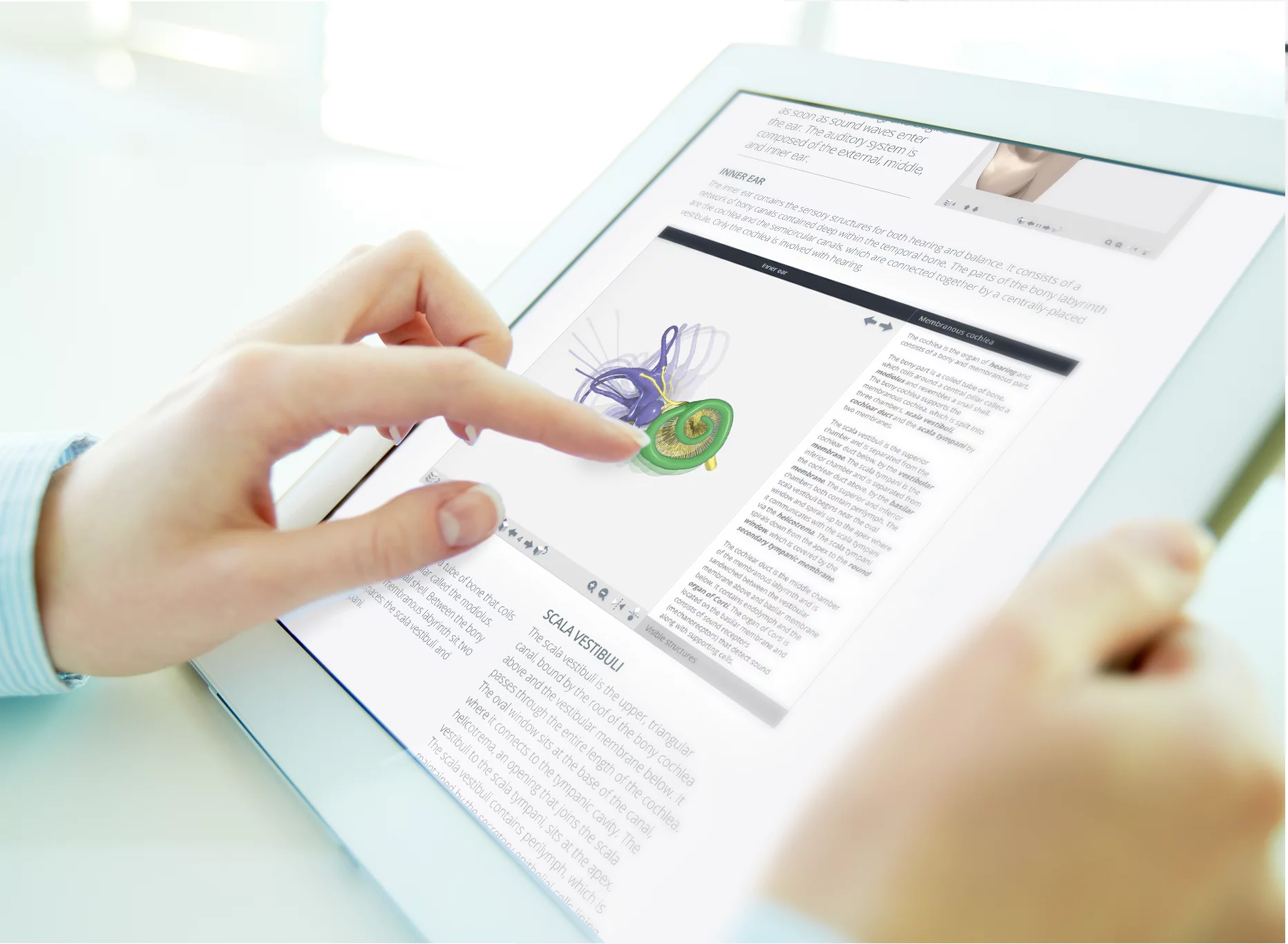
These images are most effectively seen in correlation with Primal’s trusted 3D model in our 3D Real-time content. Here you can add, remove, and view structures – in groups or in isolation – from any angle or perspective. You can even view structures through other structures, using the ‘ghost’ tool. And, as always, you get to choose whether to show the structure labeling.
You can also view these images independently in our 3D Atlas, Anatomy and Physiology, and Functional Anatomy titles.
As spatial awareness is complemented by observing diagnostic images, you will also find MRI imaging in our 3D Atlas as well as our three specialty products covering CT of the trunk and Ultrasound of the upper and lower limbs. With this array of accurate, high-quality content, you can:
- Enhance and reinforce learning during physical and virtual lab sessions on big screens or students’ personal devices.
- Let students practice or replicate dissection processes in a safe, virtual environment where the cadaveric specimen can be instantly reset to its original state – or any mistakes undone – at the touch of a button.
- Take cadaveric learning beyond the confines of physical or virtual lab sessions using resources students can access anywhere, on any device, 24/7.
Top Tips for Using Primal’s Dissection Content Inside and Outside the Lab
One of our favorite forms of feedback is hearing how you are using our digital products to support and reinforce your dissection learning. We’ve pooled your feedback with our expertise to create some top tips. These cover preparation, revision, and assessment, plus ideas for combining our digital tools with your physical specimens during in-person or virtual lab sessions themselves.
Preparation
Embed our fully labeled dissection views and slides in students’ pre-lab learning materials. This prepares students for what they can expect to see in physical specimens so they gain maximum value from the lab session itself.
Primal VR also offers a unique opportunity to dissect digital anatomy in a full VR environment, so is the perfect resource to prepare students for live cadaver lab sessions.
During Lab Sessions
Share Primal’s 3D Real-time content on large or small screens during physical or virtual lab sessions. This lets students gain a clear understanding of structures and relationships by making comparisons between how the anatomy presents in situ and what they see on screen. This can be supported further by using Primal’s imaging content.
Revision
Reinforce learning by embedding or sharing Primal’s content in post-lab materials, for students to review their learning and replicate dissection procedures. This ensures that lessons are retained.
Assessment
Assess students’ practical anatomy knowledge both online and in person using Primal’s ready-made PALMs (Perceptual and Adaptive Learning Modules) spotter-style questions or our new What Is, Where Is quiz module. Alternatively, create your own spotter-style tests using our interactive 3D views and animations, prosection slides, and dissection scenes.
Primal’s digital dissection content is the perfect fit for medical and health sciences curriculums that have access to physical cadaver specimens, as well and those that don’t. These resources are specifically designed to go beyond what the cadaver lab alone can accomplish. This means you can enhance students’ understanding of anatomy to help them achieve better outcomes.
Want to find out more? Click here to request a free trial.

