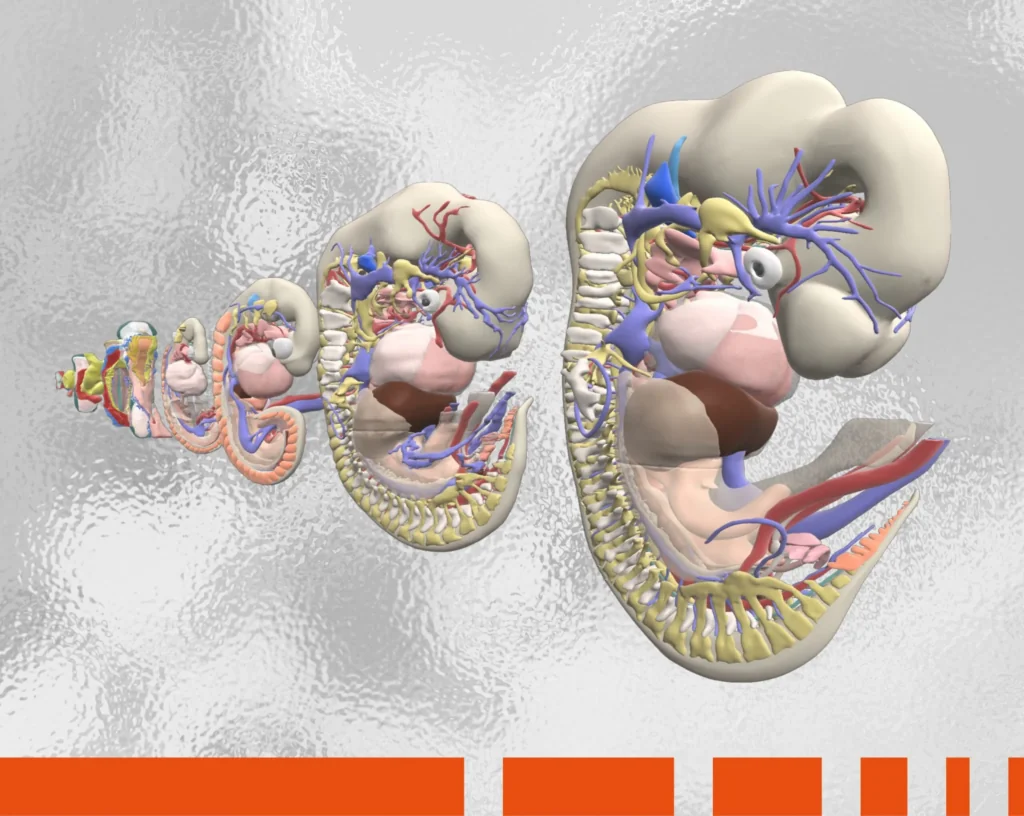
At the end of her third year Helen was required to spend a month completing a self-led project in a medicine-related subject of her choice. She used cutting-edge, 3D imagery from Primal Pictures to design an online revision tutorial for medical students based on musculoskeletal anatomy.
Primal Pictures has really helped me to design and build a high standard e-tutorial.
I was on a placement in Gloucester at the time and was keen to do a project based on musculoskeletal anatomy as I was really enjoying orthopaedics," explains Helen. "One of the orthopaedic consultants agreed to supervise my project and recommended Primal Pictures. I subsequently discovered that we had a University subscription to Primal which made the project much easier to complete.
At the University of Bristol all the anatomy teaching for medical students takes place in the first two years of their training. During this time, most of the learning takes place in the dissection room using cadavers and this is supplemented with small group sessions run by anatomy demonstrators and with lectures. Primal Pictures is now playing a role in this important training process.
“In the last year, computer-based learning has been added into anatomy teaching at Bristol,” comments Helen. “All of the teaching in the dissection room is still done in small groups with anatomy demonstrators and there are supplementary lectures to complement this learning. However, whereas we used to do one three-hour session each week; students now spend half the time in the dissection room working through computer tutorials and half the time with the demonstrators, enabling work to take place in even smaller groups. Students are also encouraged to use Primal Pictures during this computer tutorial time as well as for private study.”
Since there is no formal anatomy teaching after the second year, Helen was keen to provide an easy resource for third year students who wish to brush up on relevant practical information.
When I came to do my clinical musculoskeletal medicine placement at the end of my third year, I was feeling very rusty on my anatomy," continues Helen. "Until I sat down and did some revision I felt a bit clueless when surgeons fired questions at me in theatre. So I decided to design an e-tutorial for third year students that would be an easy and convenient means of revising relevant anatomy prior to commencing musculoskeletal medicine.
The University has an online learning environment called ‘Blackboard’ where students are able to access tutorials, most of which are designed using software called Course Genie. Helen attended teaching sessions to learn how to use this package as a platform on which to construct her e-tutorial. Since the Anatomy Department subscribes to Primal Pictures, she already had access to this software through a University login provided to her at the start of the course.
“Having completed a lot of e-tutorials in the past, I had an idea of the types that keep your attention and those that send you to sleep,” she comments. “Tutorials with blocks of text and few images are very hard to concentrate on, so wherever possible I decided to substitute images for text. As anatomy is a very visual subject, a lot of the points I wanted to make were much more easily conveyed using pictures. I also wanted to use the images to introduce interactivity to the tutorial which I hoped would further enhance its ability to hold attention and help make the information more memorable.”
Helen enjoyed the versatility of the Primal Pictures software and by incorporating the images into her project in a number of ways she was able to achieve her goals of interactivity and innovation. “Some of the images are simply used as illustrations to complement the text and some actually substitute text so the students have less reading to do. I have also integrated the images into exercises to aid learning, for example, matching a picture to the name of a bone.
“I also subscribed to another piece of software called Dragster which allowed me to create, drag and drop labels for some of the images,” continues Helen. “I have created one drag and drop image per section of the tutorial and I was pleased at how this worked out, adding a further element of interactivity.
“Primal Pictures has really helped me to design and build a high standard e-tutorial, to improve my computing skills and to broaden my knowledge and understanding of anatomy. It has also helped me to gain recognition from my peers and medical professionals as the tutorial looks impressive.”
In fact, Helen’s work has recently been nominated for a University award for innovative e-learning and she has found the software helpful in other aspects of her studies: “I’ve found Primal a really useful learning and revision tool, not only for the anatomy related to this project but also for other aspects of my clinical training. It’s much easier to grasp what’s going on, especially during surgical placements, if you have an understanding of the underlying anatomy. The interactive 3D design of the software helps apply it to real life.
I liked being able to rotate images to the desired viewpoint; building up images in layers makes the anatomy easy to understand. The accompanying text is also very useful, particularly the clinical sections. I also liked the fact that you could access an image of almost anything you wanted – the image bank is huge and I haven't seen any other resources with this volume available!
“There is no question in my mind that the end result of my e-tutorial wouldn’t have been anywhere near as good without the use of Primal Pictures. It made my tutorial stand out from the others and I feel it makes it much more educational.”
Helen achieved a 93% mark for her e-tutorial and also won the University’s Aungshuk Ghosh prize for innovative e-learning.

