Advantages of Using Digital Models
- Make the Complex Easier to Visualize and Understand
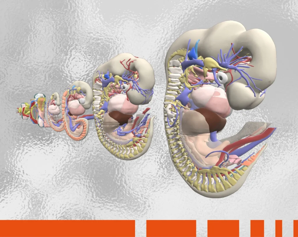
Some anatomical concepts are simply easier to visualize and grasp using a digital model. This includes changes that occur over time due to developmental stage or through disease and injury; the functional mechanics of joint movement in different positions or loads; and the physiology of cardiac muscle function.Real cadavers also have their challenges. They can be messy and cluttered, while cleaning and preserving techniques may mean that some anatomical structures don’t present well or are not readily apparent in situ. Likewise, as body donors tend to be older people, real cadavers are often diseased, which can make learning normal anatomy difficult.
- Put Students in Charge of Their Learning
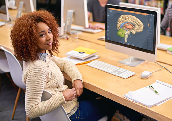
With the capacity to view anatomy from any angle, zoom in and out, highlight individual structures and virtually dissect tissue by adding and peeling away layers, digital models put students in control of their learning.This means they can reinforce their knowledge in areas where they know they are struggling, at their own pace. By immersing themselves in a 3D environment, students further their understanding of anatomical relationships and concepts to enhance their learning and knowledge retention.
- Remove the Pressure of Making Mistakes
Unlike real cadavers, digital models are resistant to degradation, damage and the dirt that comes from constant handling, meaning that the same virtual dissections can be performed time and again.What’s more, make a mistake with a digital cadaver, and you can either go back a step or two or the model can be restored to its original state, all at the click of a button. For students, this can remove the pressure of making irreversible errors and causing damage to real human tissue.
- Provide First-Rate Dissection Resources Minus the Headaches
The challenges of real cadaveric anatomy teaching and learning resources are well documented. They include financial, regulatory and practical hurdles such as rising costs, the difficulties of procuring and maintaining human specimens, and the challenges of finding physical space and/or time.There are also students who cannot perform real cadaver dissections for cultural or religious reasons. Digital cadavers are a practical alternative, offering equality of access to high-caliber learning resources, minus the headaches of the real thing.
- Access Learning 24/7 From Any Device
Most 3D digital anatomy models – including Primal’s – are optimized to be viewed on any device, including laptops, tablets and mobile phones. This makes learning flexible and repeatable, giving you instant access to content anywhere, at any time.
What Digital Dissection Doesn’t Teach
Critics highlight that digital dissection experiences are not authentic. Certainly, for many medical students, traditional physical cadaver lab marks their first encounter with a dead body. As such, the anxiety, fear – and sometimes nausea – are viewed as a rite of passage.
As students dissect a real cadaver to reveal the story – and sometimes secrets – of the individual donor, they also engage in a hands-on, tactile experience that teaches aspects of professionalism that digital programs cannot. These include empathy and respect for future patients.
Questions You Need to Ask About Digital Models
-
- How robust is the source material and can the product be trusted?
Digital 3D anatomical models can be created in a variety of ways from clinical and research imaging, surface scanning and 3D modeling. However, questions always need to be asked about the source material and whether it can be trusted.Primal’s 3D digital model is built from real CT cadaveric scans acquired from the NLM Visible Human Project. This means that all the anatomy content we create, and you experience, is both accurate and authentic.
- How robust is the source material and can the product be trusted?
- Is the Product Easy to Use?
The best eLearning tools look good and are easy to navigate and interact with. Hence, Primal’s digital design is always driven by the user. Each product is packed with compelling content, including animations, videos, slides and quizzes, that is integrated to work seamlessly in ways that you expect and are familiar with.This is not true of all digital anatomy tools, some of which may impose cognitive overload. Functionality that is not intuitive or familiar to users presents a steep learning curve that can be time-consuming and a block to engaging with the anatomy content.
Should Digital Cadavers Replace Real Ones in Future?
There is still debate about whether digital dissection can fully replace the real thing. At the end of each episode of The Anatomy Education Podcast, podcast owner, Professor James Pickering, asks each of his interviewees which they’d prefer: real cadaver dissection or an alternative.
Almost all of Pickering’s respondents and most higher-education anatomy experts generally, are reluctant to relinquish cadaveric dissection. Quite rightly, they view the experience of being able to touch, feel and see the complexities of the human body at firsthand as something that stays with students throughout their careers. But, given the choice and trusted resources, the majority would also jump at the chance of using digital dissection to compliment cadaveric anatomy.

