Industry: Creating Maximum Impact in Patient Education
For pharmaceutical and medtech companies, developing engaging and easy-to-understand patient educational materials is key in demonstrating a product’s value and purpose across its lifespan. While meeting requirements related to transparency and compliance, content needs to support healthcare professionals in promoting successful patient outcomes.
We’re here to help you achieve maximum impact depicting normal anatomy alongside that of diseases, injuries and other medical conditions. This includes images, movies, animations and diagrams for medical practitioners to use with patients in a clinical setting, and take-out materials for the person being treated to then read at home. So, if you have a medical device that corrects an anatomical defect of the heart, you can show the structure of a healthy heart next to that of an abnormal one.
Given the proliferation of specialty and more complex medicines, healthcare professionals have high expectations of the clarity and detail in any resources provided. Primal’s content, based on our 3D digital model, is derived from real scan data and covers all areas of the human body. You can customize this according to the product and the medical condition it addresses.
By providing busy medical practitioners with the best possible anatomy content to use with patients, you create strong brand awareness, trust and engagement. This includes:
- Promoting understanding of the product and the conditions it treats
- Providing context to the product’s role in treatment
- Helping healthcare professionals manage their daily workflow
- Improving patient adherence and the effectiveness of treatment plans
- Making the product offering more relevant
Click here to see an example or request a call back to discuss creating customized content that supports your treatment or product.
Higher Education: Using the Anatomy of Diseases and Injuries with Students
A winning strategy in human anatomy and physiology courses is to use medical abnormalities, pathologies and injuries to explain normal anatomical and physiological processes. All the more effective when most students are preparing for a career in the health professions, so – fingers crossed – primed to discuss diseases and disorders in class.
Choosing scenarios that students can relate to easily makes this approach even more rewarding. So, exploring the diseases, disorders, injuries and trauma of their favorite sports stars and celebrities through to examining the anatomical and physiological ramifications of obesity or gunshot and stab wounds.
Creating talking points that contrast with normal healthy anatomy and physiology lets you:
- Stimulate students’ curiosity and self-directed learning
- Support active learning with content that is inherently interesting and relevant
- Promote deeper understanding of anatomical concepts and relationships
- Explain the origin and development of anatomical structures and functions
- Develop students’ critical-thinking, problem-solving and decision-making skills
For this learning to stick, students need to be able to make comparisons between normal and abnormal anatomy using interactive visual content that is consistent. And yes, this should also be clear, accurate and from a trusted source.
Using Primal’s disease-state anatomy and physiology images, animations, movies and diagrams in your learning materials, you can spark meaningful conversations about how and why normal structure and function in the human body can deviate. This provides context to students’ learning and makes the vital connection between what they are studying and what they are likely to encounter in real life as professionals.
Contact us to explore our library of disease-state anatomy content.
Case Study: Crohn’s Disease
Crohn’s disease is a condition that can cause inflammation of any part of the digestive system. As a chronic, or lifelong, condition with periods of good health and periods of relapse and flare-ups, symptoms vary from person to person. As a result, the disease can be hard to diagnose, monitor and manage.
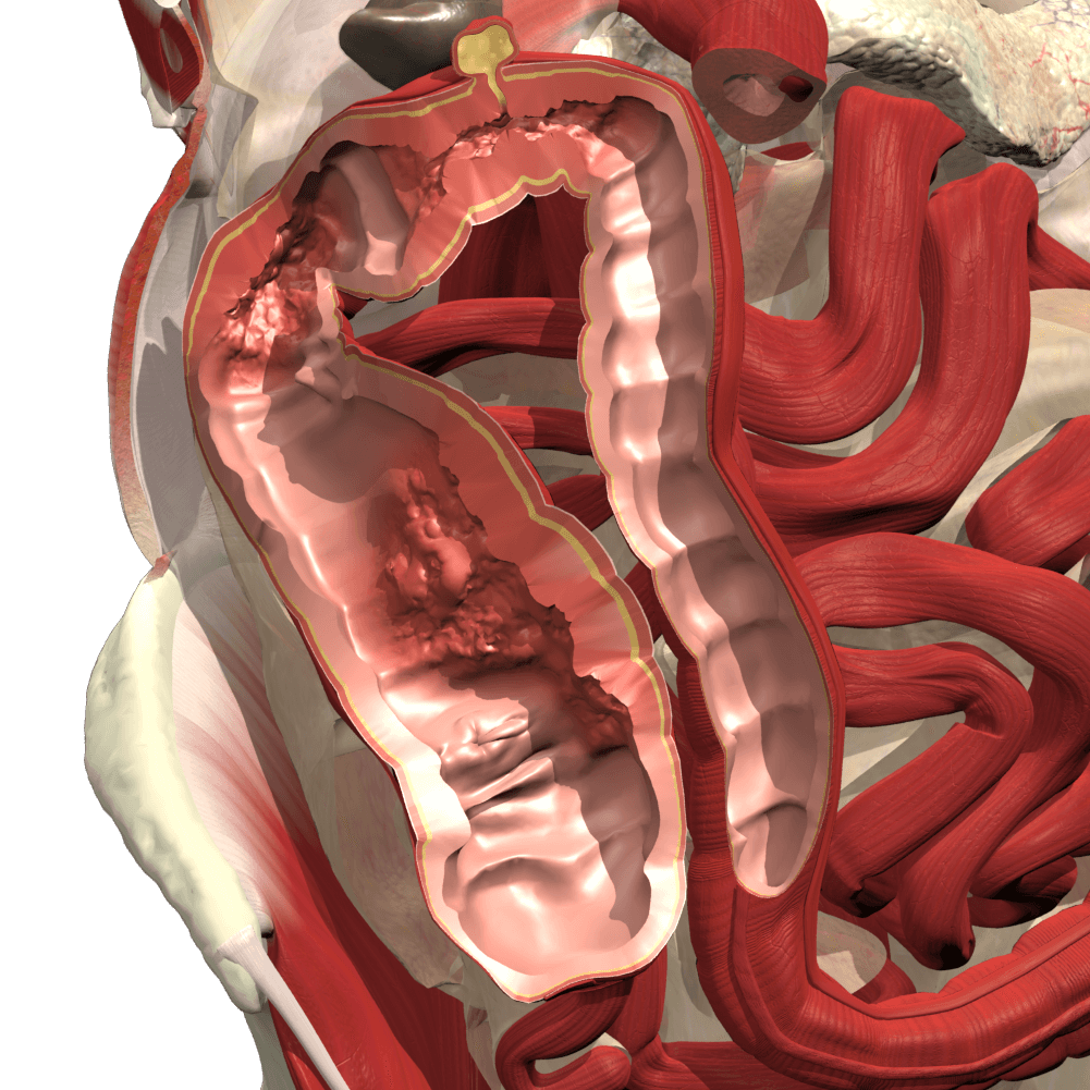
For professionals, patients, educators and students, this creates challenges that are best supported by information that is, of course, medically accurate, but also consistent, engaging and easy to grasp. So, normal and disease-damaged anatomy content that sits side by side for easy comparison of a healthy digestive system and that affected by Crohn’s is a must. See the images below showing a healthy intestine and that affected by severe strictures, abscesses and fistulas.
To illustrate and support discussions around more in-depth aspects of the condition, such as likely causes, how the disease is categorized, symptoms, and complications, we’ve included further interactive materials. These include video, animations and diagrams that detail the more intimate involvement in the disease of the individual structures through the full thickness of the intestine wall.
While a cure for Crohn’s disease may be elusive, productive doctor-patient exchanges and relationships go a long way to promote better health outcomes and make treatment less stressful. So, we’ve created a patient help sheet. This features the same visual consistency and clarity to illustrate what is happening in the body. It also provides context for the person being treated around their diagnosis and treatment plan, and the importance of following it. Healthcare professionals can share this sheet with patients for them to read at their leisure.
