Our Model
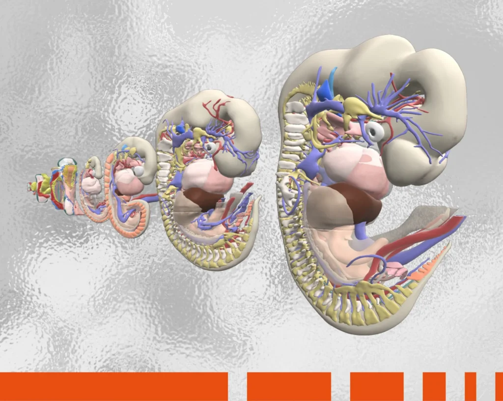
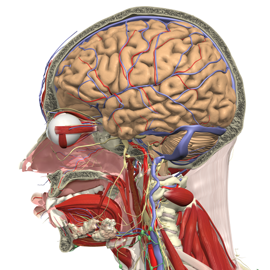
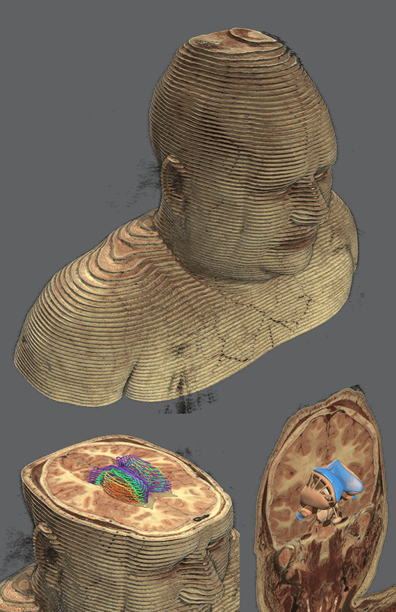
Born of a need for detailed and medically accurate 3D anatomical resources, Primal created our model from CT scans of a full human skeleton. Structures such as muscles, vessels, nerves and organs were constructed using our own MRI data and cross-sectional cadaver images from the Visible Human Project.
First, our in-house anatomy team digitally outlined every anatomical structure on the images by hand. The outlines were then used to create a 3D wireframe model of each anatomical component, ensuring the shape, position and relationships of each structure were accurate and based on real data. Next, working with our expert graphics team, they painstakingly researched the structures and details not visible in the images, so those elements could be added.
Finally, they created textures and colors to flesh out the model. Built with this level of detail, our model could be tailored to accommodate the requirements and knowledge levels of different users, from students and patients to academia, practitioners and corporate professionals. After more than 25 years, we continue to constantly refine the quality of our model today.


