12 Month Subscription
3D Real-time
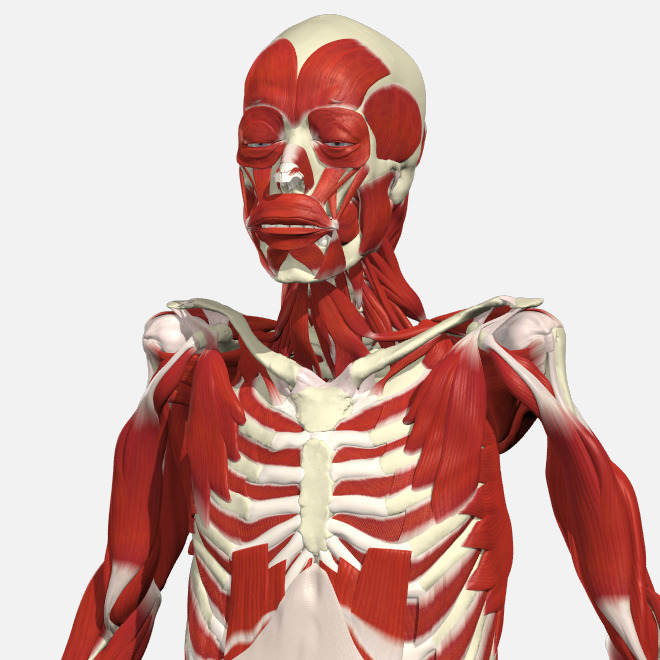
Primal’s ultimate 3D interactive anatomy experience
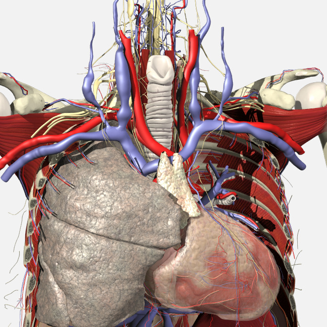
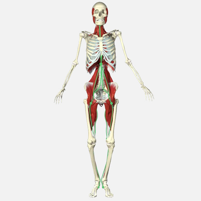
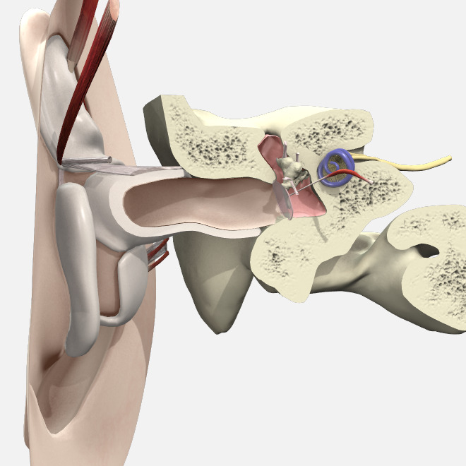
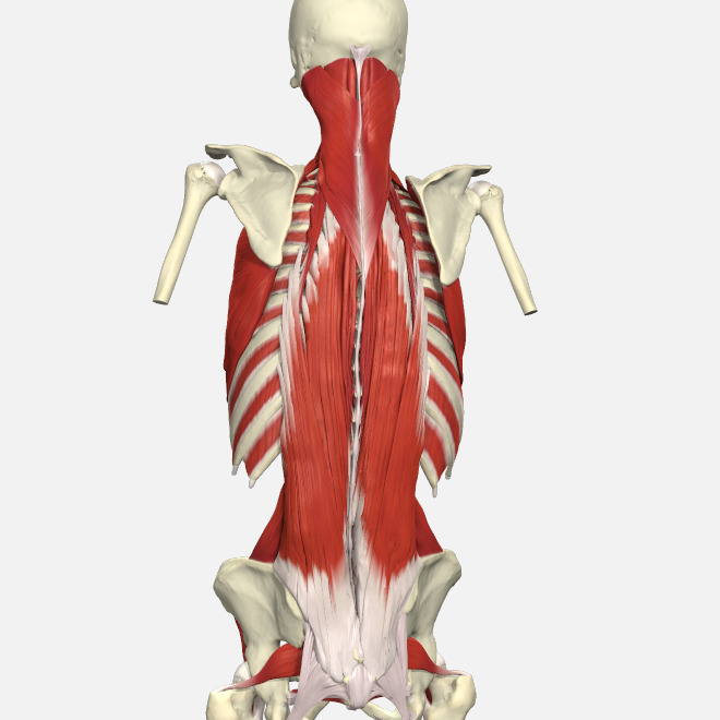
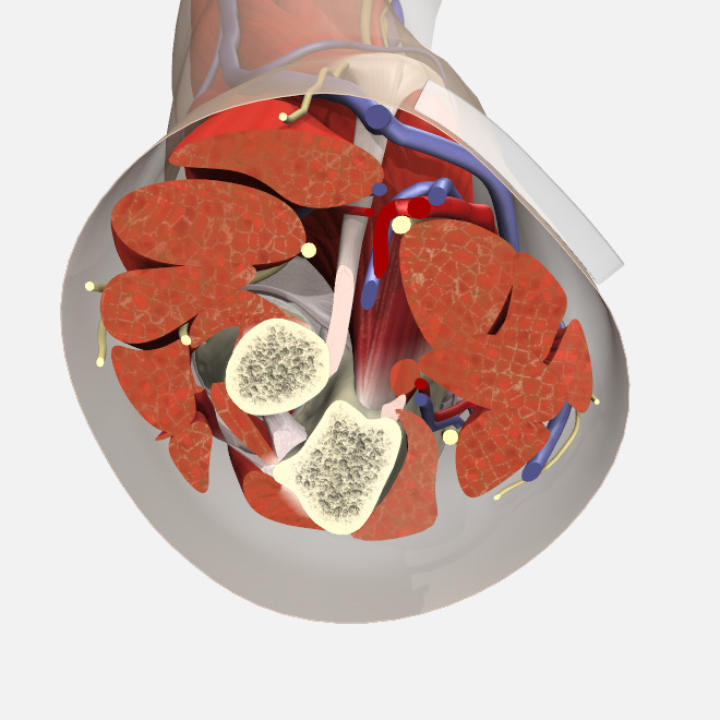
This standout 3D interactive digital anatomy resource can be used on any connected device and provides real-world context and relevance to enhance teaching, learning and communication. With a focus on digital and cadaveric dissection, it gives you ultimate control to explore the human body with unprecedented detail, accuracy and flexibility. Jump right in or customize and create content to address your specific needs – whether it’s learning anatomical relationships and detail, preparing for surgery, or educating patients.
$399.00
Included in this product

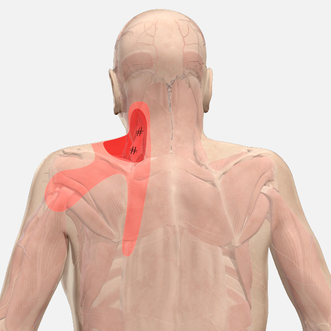
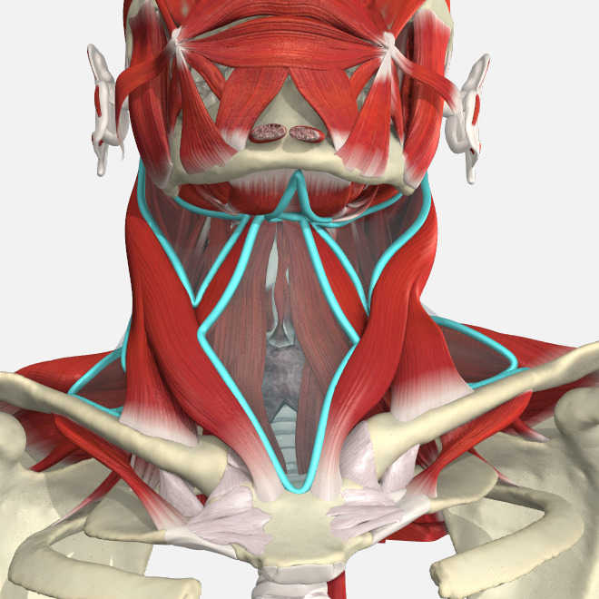
Head and Neck

Spine

Shoulder and Arm

Forearm and Hand

Thorax

Abdomen

Male Pelvis

Female Pelvis

Knee

Hip and Thigh

Leg, Ankle and Foot
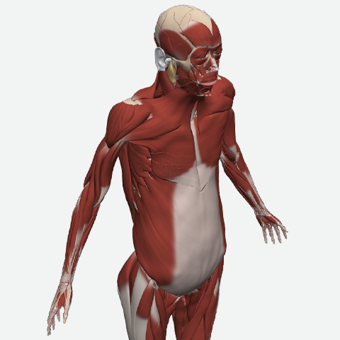
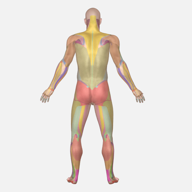
Whole Body